

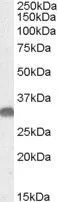
WB analysis of mouse brain lysate using GTX88681 OTUB1 antibody, Internal. Dilution : 0.05μg/ml Loading : 35μg protein in RIPA buffer
WB analysis of mouse brain lysate using GTX88681 OTUB1 antibody, Internal. Dilution : 0.05μg/ml Loading : 35μg protein in RIPA buffer
OTUB1 antibody, Internal
GTX88681
ApplicationsWestern Blot
Product group Antibodies
ReactivityHuman, Mouse
TargetOTUB1
Overview
- SupplierGeneTex
- Product NameOTUB1 antibody, Internal
- Delivery Days Customer7
- Application Supplier NoteWB: 0.03-0.1microg/ml. *Optimal dilutions/concentrations should be determined by the researcher.Not tested in other applications.
- ApplicationsWestern Blot
- CertificationResearch Use Only
- ClonalityPolyclonal
- Concentration0.50 mg/ml
- ConjugateUnconjugated
- Gene ID55611
- Target nameOTUB1
- Target descriptionOTU deubiquitinase, ubiquitin aldehyde binding 1
- Target synonymsHSPC263, OTB1, OTU1, ubiquitin thioesterase OTUB1, OTU domain, ubiquitin aldehyde binding 1, OTU domain-containing ubiquitin aldehyde-binding protein 1, OTU-domain Ubal-binding 1, deubiquitinating enzyme OTUB1, otubain-1, ubiquitin-specific protease otubain 1, ubiquitin-specific-processing protease OTUB1
- HostGoat
- IsotypeIgG
- Protein IDQ96FW1
- Protein NameUbiquitin thioesterase OTUB1
- Scientific DescriptionThe product of this gene is a member of the OTU (ovarian tumor) superfamily of predicted cysteine proteases. The encoded protein is a highly specific ubiquitin iso-peptidase, and cleaves ubiquitin from branched poly-ubiquitin chains but not from ubiquitinated substrates. It interacts with another ubiquitin protease and an E3 ubiquitin ligase that inhibits cytokine gene transcription in the immune system. It is proposed to function in specific ubiquitin-dependent pathways, possibly by providing an editing function for polyubiquitin chain growth. Alternative splicing results in multiple transcript variants. [provided by RefSeq, Jul 2008]
- ReactivityHuman, Mouse
- Storage Instruction-20°C or -80°C,2°C to 8°C
- UNSPSC41116161
Datasheet
Related products
Product group Antibodies
Anti-OTUB1 AntibodyA16987
ApplicationsWestern Blot
ReactivityHuman, Mouse
- SizePrice

Product group Antibodies
Anti-OTUB1 Antibody Picoband(r)A04361-1-CARRIER-FREE
ApplicationsFlow Cytometry, Western Blot, ELISA, ImmunoHistoChemistry
ReactivityHuman, Mouse, Rat
TargetOTUB1
- SizePrice
Product group Antibodies
Anti-OTUB1 Antibody144-10313
ApplicationsWestern Blot
ReactivityHuman, Mouse
TargetOTUB1
- SizePrice
Product group Antibodies
OTUB1 Polyclonal AntibodyBS-11229R
ApplicationsImmunoFluorescence, Western Blot, ELISA, ImmunoCytoChemistry, ImmunoHistoChemistry, ImmunoHistoChemistry Frozen, ImmunoHistoChemistry Paraffin
- SizePrice
Product group Antibodies

Goat anti-OTUB1EB08653
ApplicationsWestern Blot, ELISA
ReactivityBovine, Human, Mouse, Rat
TargetOTUB1
- SizePrice
Product group Antibodies
OTUB1 Polyclonal AntibodyCAC14045
ApplicationsWestern Blot, ELISA, ImmunoHistoChemistry
TargetOTUB1
- SizePrice
Product group Antibodies
OTUB1 AntibodyCSB-PA03595A0RB
ApplicationsWestern Blot, ELISA, ImmunoHistoChemistry
ReactivityHuman
TargetOTUB1
- SizePrice
Product group Antibodies

OTUB1 / OTU1 AntibodyLS-C401910
ApplicationsWestern Blot, ELISA
ReactivityHuman, Mouse, Rat
TargetOTUB1
- SizePrice
![Various whole cell extracts (30 μg) were separated by 12% SDS-PAGE, and the membrane was blotted with OTUB1 antibody [N1C1] (GTX101973) diluted at 1:1000. The HRP-conjugated anti-rabbit IgG antibody (GTX213110-01) was used to detect the primary antibody, and the signal was developed with Trident ECL plus-Enhanced.](https://www.genetex.com/upload/website/prouct_img/normal/GTX101973/GTX101973_39694_20220930_WB_H_M_R_22101319_150.webp)
Product group Antibodies
OTUB1 antibody [N1C1]GTX101973
ApplicationsImmunoFluorescence, Western Blot, ImmunoCytoChemistry, ImmunoHistoChemistry, ImmunoHistoChemistry Paraffin
ReactivityHuman, Mouse, Rat
TargetOTUB1
- SizePrice

Product group Antibodies
Anti-OTUB1 AntibodyHPA039176
ApplicationsWestern Blot, ImmunoHistoChemistry
ReactivityHuman, Mouse, Rat
TargetOTUB1
- SizePrice