

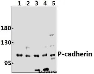
WB analysis of various samples using GTX66687 P-Cadherin antibody. Lane1 : A375 whole cell lysate(40ug)
Lane2 : C6 whole cell lysate(40ug)
Lane3 : Beas-2B whole cell lysate(40ug)
Lane4 : EC9706 whole cell lysate(40ug)
Lane5 : CT26 whole cell lysate(40ug) Dilution : 1:500
WB analysis of various samples using GTX66687 P-Cadherin antibody. Lane1 : A375 whole cell lysate(40ug)
Lane2 : C6 whole cell lysate(40ug)
Lane3 : Beas-2B whole cell lysate(40ug)
Lane4 : EC9706 whole cell lysate(40ug)
Lane5 : CT26 whole cell lysate(40ug) Dilution : 1:500
P-Cadherin antibody
GTX66687
ApplicationsWestern Blot
Product group Antibodies
ReactivityHuman, Mouse, Rat
TargetCDH3
Overview
- SupplierGeneTex
- Product NameP-Cadherin antibody
- Delivery Days Customer9
- Application Supplier NoteWB: 1:500-1:1000. *Optimal dilutions/concentrations should be determined by the researcher.Not tested in other applications.
- ApplicationsWestern Blot
- CertificationResearch Use Only
- ClonalityPolyclonal
- Concentration1 mg/ml
- ConjugateUnconjugated
- Gene ID1001
- Target nameCDH3
- Target descriptioncadherin 3
- Target synonymsCDHP, HJMD, PCAD, cadherin-3, cadherin 3, type 1, P-cadherin (placental), calcium-dependent adhesion protein, placental
- HostRabbit
- IsotypeIgG
- Protein IDP22223
- Protein NameCadherin-3
- Scientific DescriptionThis gene encodes a classical cadherin of the cadherin superfamily. Alternative splicing results in multiple transcript variants, at least one of which encodes a preproprotein that is proteolytically processed to generate the mature glycoprotein. This calcium-dependent cell-cell adhesion protein is comprised of five extracellular cadherin repeats, a transmembrane region and a highly conserved cytoplasmic tail. This gene is located in a gene cluster in a region on the long arm of chromosome 16 that is involved in loss of heterozygosity events in breast and prostate cancer. In addition, aberrant expression of this protein is observed in cervical adenocarcinomas. Mutations in this gene are associated with hypotrichosis with juvenile macular dystrophy and ectodermal dysplasia, ectrodactyly, and macular dystrophy syndrome (EEMS). [provided by RefSeq, Nov 2015]
- ReactivityHuman, Mouse, Rat
- Storage Instruction-20°C or -80°C,2°C to 8°C
- UNSPSC12352203
Datasheet
Related products
Product group Antibodies

Anti-P-Cadherin [6A9]AB02535-1.1
ApplicationsImmunoFluorescence, ImmunoPrecipitation, Western Blot, ImmunoHistoChemistry
ReactivityHuman
TargetCDH3
- SizePrice
Product group Antibodies
Anti-CDH3 Antibody144-63143
ApplicationsWestern Blot, ImmunoHistoChemistry
ReactivityHuman, Mouse, Rat
TargetCDH3
- SizePrice

Product group Antibodies
Anti-P-Cadherin-3 CDH3-Antibody Picoband(r)A03353-1-CARRIER-FREE
ApplicationsFlow Cytometry, ImmunoFluorescence, Western Blot, ELISA, ImmunoCytoChemistry
ReactivityHuman
TargetCDH3
- SizePrice
Product group Antibodies
References
P-Cadherin antibodyGTX113648
ApplicationsWestern Blot, ImmunoHistoChemistry, ImmunoHistoChemistry Paraffin
ReactivityHuman, Mouse
TargetCDH3
- SizePrice
Product group Antibodies
P-Cadherin (N-terminal region) AntibodyBSM-70369M
ApplicationsWestern Blot
ReactivityHuman, Mouse, Rat
TargetCDH3
- SizePrice
Product group Antibodies
Anti-CDH3 AntibodyA99919
ApplicationsWestern Blot, ELISA, ImmunoHistoChemistry
ReactivityHuman
- SizePrice
![ICC/IF analysis of BEAS-2B Cells using GTX19350 P-Cadherin antibody [6A9]. Cells were probed without (right) or with(left) an antibody. Green : Primary antibody Blue : Nuclei Red : Actin Fixation : formaldehyde Dilution : 1:20 overnight at 4oC](https://www.genetex.com/upload/website/prouct_img/normal/GTX19350/GTX19350_366_ICC-IF_w_23060620_354.webp)
Product group Antibodies
P-Cadherin antibody [6A9]GTX19350
ApplicationsImmunoFluorescence, ImmunoPrecipitation, Western Blot, ImmunoCytoChemistry, Neutralisation/Blocking
ReactivityHuman, Mouse
TargetCDH3
- SizePrice
Product group Antibodies
ApplicationsWestern Blot, ELISA, ImmunoCytoChemistry, ImmunoHistoChemistry, ImmunoHistoChemistry Frozen, ImmunoHistoChemistry Paraffin
ReactivityMouse
TargetCDH3
- SizePrice
Product group Antibodies
ApplicationsWestern Blot, ImmunoHistoChemistry, ImmunoHistoChemistry Frozen, Neutralisation/Blocking
ReactivityHuman
TargetCDH3
- SizePrice