

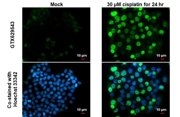
p21 Cip1 antibody [GT1032] detects p21 Cip1 protein at nucleus by immunofluorescent analysis. Sample: Mock and treated HCT116 cells were fixed in 4% paraformaldehyde at RT for 15 min. Green: p21 Cip1 stained by p21 Cip1 antibody [GT1032] (GTX629543) diluted at 1:500. Blue: Hoechst 33342 staining. Scale bar= 10 μm.
![p21 Cip1 antibody [GT1032] detects p21 Cip1 protein at nucleus by immunofluorescent analysis. Sample: MCF7 cells were fixed in 4% paraformaldehyde at RT for 15 min. Green: CDK4 protein stained by CDK4 antibody (GTX102993) diluted at 1:1000. Red: p21 Cip1 protein stained by p21 Cip1 antibody [GT1032] (GTX629543) diluted at 1:500. Blue: Hoechst 33342 staining.](https://www.genetex.com/upload/website/prouct_img/normal/GTX629543/GTX629543_41365_20150518_IFA_w_23061202_449.webp "p21 Cip1 antibody [GT1032] detects p21 Cip1 protein at nucleus by immunofluorescent analysis. Sample: MCF7 cells were fixed in 4% paraformaldehyde at RT for 15 min. Green: CDK4 protein stained by CDK4 antibody (GTX102993) diluted at 1:1000. Red: p21 Cip1 protein stained by p21 Cip1 antibody [GT1032] (GTX629543) diluted at 1:500. Blue: Hoechst 33342 staining.")
![p21 Cip1 antibody [GT1032] immunoprecipitates CDKN1A protein in IP experiments. IP samples: HCT-116 whole cell extract treat with 30uM cisplatin for 48 hr A. 30 μg HCT-116 whole cell extract treat with 30uM cisplatin for 48 hr B. Control with 4 μg of preimmune Mouse IgG C. Immunoprecipitation of CDKN1A protein by 4 μg p21 Cip1 antibody [GT1032] (GTX629543) 15 % SDS-PAGE The immunoprecipitated CDKN1A protein was detected by p21 Cip1 antibody [GT1032] (GTX629543) diluted at 1:1000. [EasyBlot anti-mouse IgG (GTX221667-01) was used as a secondary reagent]](https://www.genetex.com/upload/website/prouct_img/normal/GTX629543/GTX629543_41365_IP_w_23061202_810.webp "p21 Cip1 antibody [GT1032] immunoprecipitates CDKN1A protein in IP experiments. IP samples: HCT-116 whole cell extract treat with 30uM cisplatin for 48 hr A. 30 μg HCT-116 whole cell extract treat with 30uM cisplatin for 48 hr B. Control with 4 μg of preimmune Mouse IgG C. Immunoprecipitation of CDKN1A protein by 4 μg p21 Cip1 antibody [GT1032] (GTX629543) 15 % SDS-PAGE The immunoprecipitated CDKN1A protein was detected by p21 Cip1 antibody [GT1032] (GTX629543) diluted at 1:1000. [EasyBlot anti-mouse IgG (GTX221667-01) was used as a secondary reagent]")
![p21 Cip1 antibody [GT1032] (GTX629543) detects p21 Cip1 by flow cytometry analysis. Sample: HepG2 cell. Black: Unlabelled sample was used as a control. Red: p21 Cip1 antibody [GT1032] (GTX629543) dilution: 1:50. Acquisition of 20,000 events were collected for FACS analysis.](https://www.genetex.com/upload/website/prouct_img/normal/GTX629543/GTX629543_41939_20200319_FACS_w_23061202_349.webp "p21 Cip1 antibody [GT1032] (GTX629543) detects p21 Cip1 by flow cytometry analysis. Sample: HepG2 cell. Black: Unlabelled sample was used as a control. Red: p21 Cip1 antibody [GT1032] (GTX629543) dilution: 1:50. Acquisition of 20,000 events were collected for FACS analysis.")
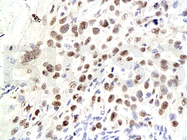
![p21 Cip1 antibody [GT1032] detects p21 Cip1 protein at nucleus by immunohistochemical analysis. Sample: Paraffin-embedded human lung cancer. p21 Cip1 stained by p21 Cip1 antibody [GT1032] (GTX629543) diluted at 1:200. Antigen Retrieval: Citrate buffer, pH 6.0, 15 min](https://www.genetex.com/upload/website/prouct_img/normal/GTX629543/GTX629543_43444_20200619_IHC-P_w_23061202_107.webp "p21 Cip1 antibody [GT1032] detects p21 Cip1 protein at nucleus by immunohistochemical analysis. Sample: Paraffin-embedded human lung cancer. p21 Cip1 stained by p21 Cip1 antibody [GT1032] (GTX629543) diluted at 1:200. Antigen Retrieval: Citrate buffer, pH 6.0, 15 min")


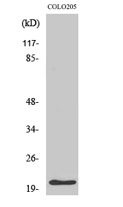
![Untreated (–) and treated (+) HCT-116 whole cell extracts (30 μg) were separated by 15% SDS-PAGE, and the membrane was blotted with p21 Cip1 antibody [GT1032] (GTX629543) diluted at 1:5000. The HRP-conjugated anti-mouse IgG antibody (GTX213111-01) was used to detect the primary antibody.](https://www.genetex.com/upload/website/prouct_img/normal/GTX629543/GTX629543_43444_20190118_WB_treatment_Cisplatin_25030600_406.webp "Untreated (–) and treated (+) HCT-116 whole cell extracts (30 μg) were separated by 15% SDS-PAGE, and the membrane was blotted with p21 Cip1 antibody [GT1032] (GTX629543) diluted at 1:5000. The HRP-conjugated anti-mouse IgG antibody (GTX213111-01) was used to detect the primary antibody.")
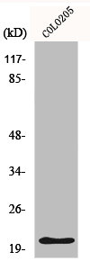
![Wild-type (WT) and p21 Cip1 knockout (KO) HeLa cell extracts (30 μg) were separated by 15% SDS-PAGE, and the membrane was blotted with p21 Cip1 antibody [GT1032] (GTX629543) diluted at 1:1000. The HRP-conjugated anti-mouse IgG antibody (GTX213111-01) was used to detect the primary antibody, and the signal was developed with Trident ECL plus-Enhanced.](https://www.genetex.com/upload/website/prouct_img/normal/GTX629543/GTX629543_43444_20190607_WB_KO_watermark_25030600_767.webp "Wild-type (WT) and p21 Cip1 knockout (KO) HeLa cell extracts (30 μg) were separated by 15% SDS-PAGE, and the membrane was blotted with p21 Cip1 antibody [GT1032] (GTX629543) diluted at 1:1000. The HRP-conjugated anti-mouse IgG antibody (GTX213111-01) was used to detect the primary antibody, and the signal was developed with Trident ECL plus-Enhanced.")
p21 Cip1 antibody [GT1032] detects p21 Cip1 protein at nucleus by immunofluorescent analysis. Sample: Mock and treated HCT116 cells were fixed in 4% paraformaldehyde at RT for 15 min. Green: p21 Cip1 stained by p21 Cip1 antibody [GT1032] (GTX629543) diluted at 1:500. Blue: Hoechst 33342 staining. Scale bar= 10 μm.
p21 Cip1 antibody [GT1032]
GTX629543
ApplicationsFlow Cytometry, ImmunoFluorescence, ImmunoPrecipitation, Western Blot, ImmunoCytoChemistry, ImmunoHistoChemistry, ImmunoHistoChemistry Paraffin
Product group Antibodies
ReactivityHuman, Mouse
TargetCDKN1A
Overview
- SupplierGeneTex
- Product Namep21 Cip1 antibody [GT1032]
- Delivery Days Customer9
- Application Supplier NoteWB: 1:500-1:3000. ICC/IF: 1:100-1:1000. IP: 1:100-1:500. *Optimal dilutions/concentrations should be determined by the researcher.Not tested in other applications.
- ApplicationsFlow Cytometry, ImmunoFluorescence, ImmunoPrecipitation, Western Blot, ImmunoCytoChemistry, ImmunoHistoChemistry, ImmunoHistoChemistry Paraffin
- CertificationResearch Use Only
- ClonalityMonoclonal
- Clone IDGT1032
- Concentration0.8 mg/ml
- ConjugateUnconjugated
- Gene ID1026
- Target nameCDKN1A
- Target descriptioncyclin dependent kinase inhibitor 1A
- Target synonymsCAP20, CDKN1, CIP1, MDA-6, P21, SDI1, WAF1, p21CIP1, cyclin-dependent kinase inhibitor 1, CDK-interacting protein 1, CDK-interaction protein 1, DNA synthesis inhibitor, cyclin-dependent kinase inhibitor 1A (p21, Cip1), melanoma differentiation associated protein 6, wild-type p53-activated fragment 1
- HostMouse
- IsotypeIgG2a
- Protein IDP38936
- Protein NameCyclin-dependent kinase inhibitor 1
- Scientific DescriptionThis gene encodes a potent cyclin-dependent kinase inhibitor. The encoded protein binds to and inhibits the activity of cyclin-CDK2 or -CDK4 complexes, and thus functions as a regulator of cell cycle progression at G1. The expression of this gene is tightly controlled by the tumor suppressor protein p53, through which this protein mediates the p53-dependent cell cycle G1 phase arrest in response to a variety of stress stimuli. This protein can interact with proliferating cell nuclear antigen (PCNA), a DNA polymerase accessory factor, and plays a regulatory role in S phase DNA replication and DNA damage repair. This protein was reported to be specifically cleaved by CASP3-like caspases, which thus leads to a dramatic activation of CDK2, and may be instrumental in the execution of apoptosis following caspase activation. Two alternatively spliced variants, which encode an identical protein, have been reported. [provided by RefSeq]
- ReactivityHuman, Mouse
- Storage Instruction-20°C or -80°C,2°C to 8°C
- UNSPSC41116161
Datasheet
Related products
Product group Antibodies
Anti-p21 Antibody130-10051
ApplicationsELISA
ReactivityHuman
TargetCDKN1A
- SizePrice
Product group Antibodies

P21Cip1 AntibodyABX034318
ApplicationsImmunoFluorescence, Western Blot, ELISA, ImmunoCytoChemistry
- SizePrice

Product group Antibodies
Anti-CDKN1A AntibodyAMAB91832
ApplicationsWestern Blot, ImmunoCytoChemistry
ReactivityHuman
TargetCDKN1A
- SizePrice
Product group Antibodies
Anti-p21 AntibodyA40795
ApplicationsWestern Blot, ELISA
ReactivityHuman, Mouse, Rat
- SizePrice

Product group Antibodies
Anti-P21/CDKN1A Antibody Picoband(r)A00145-1-CARRIER-FREE
ApplicationsImmunoFluorescence, Western Blot, ELISA, ImmunoCytoChemistry, ImmunoHistoChemistry
ReactivityHuman
TargetCDKN1A
- SizePrice
Product group Antibodies
References
CDKN1A/p21 Polyclonal AntibodyBS-0741R
ApplicationsImmunoFluorescence, Western Blot, ELISA, ImmunoCytoChemistry, ImmunoHistoChemistry, ImmunoHistoChemistry Frozen, ImmunoHistoChemistry Paraffin
ReactivityBovine, Canine, Chicken, Human, Mouse, Rat
TargetCDKN1A
- SizePrice
Product group Antibodies
CDKN1A AntibodyCSB-PA003623
ApplicationsWestern Blot, ELISA
ReactivityHuman, Mouse, Rat
TargetCDKN1A
- SizePrice
Product group Antibodies
Cdkn1A Polyclonal AntibodyCAC08304
ApplicationsImmunoFluorescence, ELISA, ImmunoHistoChemistry
TargetCDKN1A
- SizePrice
Product group Antibodies
p21 Cip1 antibodyGTX135142
ApplicationsWestern Blot
ReactivityHuman
TargetCDKN1A
- SizePrice