

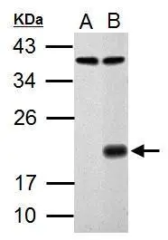
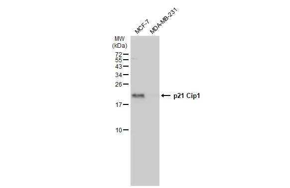
Sample (30 μg of whole cell lysate) A: HCT116 B: HCT116 treated with 30 μM cisplatin for 24 hr 12% SDS PAGE GTX100444 diluted at 1:1000 The HRP-conjugated anti-rabbit IgG antibody (GTX213110-01) was used to detect the primary antibody.
![p21 Cip1 antibody detects p21 Cip1 protein at nucleus by immunofluorescent analysis. Samples: HCT116 cells mock (left) and treated with 30 μM Cisplatin for 24 hrs (right) were fixed in 4% paraformaldehyde at RT for 15 min. Green: p21 Cip1 protein stained by p21 Cip1 antibody (GTX100444) diluted at 1:1000. Red: Histone H3S10ph (phospho Ser10), a nucleus marker, stained by Histone H3S10ph (phospho Ser10) antibody [GT921] (GTX630185) diluted at 1:500. Blue: Hoechst 33342 staining.](https://www.genetex.com/upload/website/prouct_img/normal/GTX100444/GTX100444_40996_20150518_IFA_w_23060100_609.webp "p21 Cip1 antibody detects p21 Cip1 protein at nucleus by immunofluorescent analysis. Samples: HCT116 cells mock (left) and treated with 30 μM Cisplatin for 24 hrs (right) were fixed in 4% paraformaldehyde at RT for 15 min. Green: p21 Cip1 protein stained by p21 Cip1 antibody (GTX100444) diluted at 1:1000. Red: Histone H3S10ph (phospho Ser10), a nucleus marker, stained by Histone H3S10ph (phospho Ser10) antibody [GT921] (GTX630185) diluted at 1:500. Blue: Hoechst 33342 staining.")

Sample (30 μg of whole cell lysate) A: HCT116 B: HCT116 treated with 30 μM cisplatin for 24 hr 12% SDS PAGE GTX100444 diluted at 1:1000 The HRP-conjugated anti-rabbit IgG antibody (GTX213110-01) was used to detect the primary antibody.
p21 Cip1 antibody
GTX100444
ApplicationsImmunoFluorescence, Western Blot, ImmunoCytoChemistry
Product group Antibodies
ReactivityHuman, Rat
TargetCDKN1A
Overview
- SupplierGeneTex
- Product Namep21 Cip1 antibody
- Delivery Days Customer9
- Application Supplier NoteWB: 1:500-1:3000. ICC/IF: 1:100-1:1000. *Optimal dilutions/concentrations should be determined by the researcher.Not tested in other applications.
- ApplicationsImmunoFluorescence, Western Blot, ImmunoCytoChemistry
- CertificationResearch Use Only
- ClonalityPolyclonal
- Concentration1 mg/ml
- ConjugateUnconjugated
- Gene ID1026
- Target nameCDKN1A
- Target descriptioncyclin dependent kinase inhibitor 1A
- Target synonymsCAP20, CDKN1, CIP1, MDA-6, P21, SDI1, WAF1, p21CIP1, cyclin-dependent kinase inhibitor 1, CDK-interacting protein 1, CDK-interaction protein 1, DNA synthesis inhibitor, cyclin-dependent kinase inhibitor 1A (p21, Cip1), melanoma differentiation associated protein 6, wild-type p53-activated fragment 1
- HostRabbit
- IsotypeIgG
- Protein IDP38936
- Protein NameCyclin-dependent kinase inhibitor 1
- Scientific DescriptionThis gene encodes a potent cyclin-dependent kinase inhibitor. The encoded protein binds to and inhibits the activity of cyclin-CDK2 or -CDK4 complexes, and thus functions as a regulator of cell cycle progression at G1. The expression of this gene is tightly controlled by the tumor suppressor protein p53, through which this protein mediates the p53-dependent cell cycle G1 phase arrest in response to a variety of stress stimuli. This protein can interact with proliferating cell nuclear antigen (PCNA), a DNA polymerase accessory factor, and plays a regulatory role in S phase DNA replication and DNA damage repair. This protein was reported to be specifically cleaved by CASP3-like caspases, which thus leads to a dramatic activation of CDK2, and may be instrumental in the execution of apoptosis following caspase activation. Two alternatively spliced variants, which encode an identical protein, have been reported. [provided by RefSeq]
- ReactivityHuman, Rat
- Storage Instruction-20°C or -80°C,2°C to 8°C
- UNSPSC41116161
Datasheet
Related products
Product group Antibodies
Anti-p21 Antibody130-10051
ApplicationsELISA
ReactivityHuman
TargetCDKN1A
- SizePrice
Product group Antibodies

P21Cip1 AntibodyABX034318
ApplicationsImmunoFluorescence, Western Blot, ELISA, ImmunoCytoChemistry
- SizePrice

Product group Antibodies
Anti-CDKN1A AntibodyAMAB91832
ApplicationsWestern Blot, ImmunoCytoChemistry
ReactivityHuman
TargetCDKN1A
- SizePrice
Product group Antibodies
Anti-p21 AntibodyA40795
ApplicationsWestern Blot, ELISA
ReactivityHuman, Mouse, Rat
- SizePrice

Product group Antibodies
Anti-P21/CDKN1A Antibody Picoband(r)A00145-1-CARRIER-FREE
ApplicationsImmunoFluorescence, Western Blot, ELISA, ImmunoCytoChemistry, ImmunoHistoChemistry
ReactivityHuman
TargetCDKN1A
- SizePrice
Product group Antibodies
References
CDKN1A/p21 Polyclonal AntibodyBS-0741R
ApplicationsImmunoFluorescence, Western Blot, ELISA, ImmunoCytoChemistry, ImmunoHistoChemistry, ImmunoHistoChemistry Frozen, ImmunoHistoChemistry Paraffin
ReactivityBovine, Canine, Chicken, Human, Mouse, Rat
TargetCDKN1A
- SizePrice
Product group Antibodies
CDKN1A AntibodyCSB-PA003623
ApplicationsWestern Blot, ELISA
ReactivityHuman, Mouse, Rat
TargetCDKN1A
- SizePrice
Product group Antibodies
Cdkn1A Polyclonal AntibodyCAC08304
ApplicationsImmunoFluorescence, ELISA, ImmunoHistoChemistry
TargetCDKN1A
- SizePrice
Product group Antibodies
p21 Cip1 antibodyGTX135142
ApplicationsWestern Blot
ReactivityHuman
TargetCDKN1A
- SizePrice