

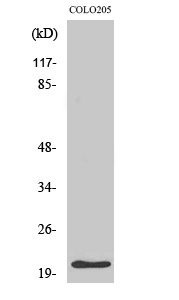
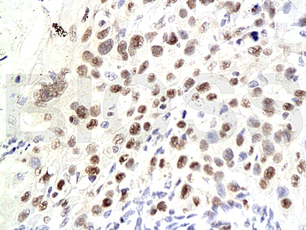
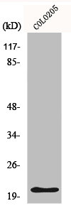
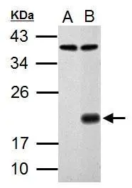
P21Cip1 Antibody
ABX034318
ApplicationsImmunoFluorescence, Western Blot, ELISA, ImmunoCytoChemistry
Product group Antibodies
Overview
- SupplierAbbexa
- Product NameP21Cip1 Antibody
- Delivery Days Customer12
- ApplicationsImmunoFluorescence, Western Blot, ELISA, ImmunoCytoChemistry
- CertificationResearch Use Only
- ClonalityPolyclonal
- ConjugateUnconjugated
- HostRabbit
- UNSPSC12352203
Related products
Product group Antibodies
Anti-p21 Antibody130-10051
ApplicationsELISA
ReactivityHuman
TargetCDKN1A
- SizePrice

Product group Antibodies
Anti-CDKN1A AntibodyAMAB91832
ApplicationsWestern Blot, ImmunoCytoChemistry
ReactivityHuman
TargetCDKN1A
- SizePrice
Product group Antibodies
Anti-p21 AntibodyA40795
ApplicationsWestern Blot, ELISA
ReactivityHuman, Mouse, Rat
- SizePrice

Product group Antibodies
Anti-P21/CDKN1A Antibody Picoband(r)A00145-1-CARRIER-FREE
ApplicationsImmunoFluorescence, Western Blot, ELISA, ImmunoCytoChemistry, ImmunoHistoChemistry
ReactivityHuman
TargetCDKN1A
- SizePrice
Product group Antibodies
References
CDKN1A/p21 Polyclonal AntibodyBS-0741R
ApplicationsImmunoFluorescence, Western Blot, ELISA, ImmunoCytoChemistry, ImmunoHistoChemistry, ImmunoHistoChemistry Frozen, ImmunoHistoChemistry Paraffin
ReactivityBovine, Canine, Chicken, Human, Mouse, Rat
TargetCDKN1A
- SizePrice
Product group Antibodies
CDKN1A AntibodyCSB-PA003623
ApplicationsWestern Blot, ELISA
ReactivityHuman, Mouse, Rat
TargetCDKN1A
- SizePrice
Product group Antibodies
Cdkn1A Polyclonal AntibodyCAC08304
ApplicationsImmunoFluorescence, ELISA, ImmunoHistoChemistry
TargetCDKN1A
- SizePrice
Product group Antibodies

ApplicationsELISA
ReactivityHuman
TargetCDKN1A
- SizePrice
Product group Antibodies
p21 Cip1 antibodyGTX100444
ApplicationsImmunoFluorescence, Western Blot, ImmunoCytoChemistry
ReactivityHuman, Rat
TargetCDKN1A
- SizePrice