

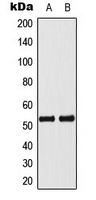
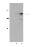
WB analysis of Actinomycin-treated MCF7 (A), Jurkat (B) whole cell lysates using GTX32360 p53 (phospho Ser33) antibody.
WB analysis of Actinomycin-treated MCF7 (A), Jurkat (B) whole cell lysates using GTX32360 p53 (phospho Ser33) antibody.
p53 (phospho Ser33) antibody
GTX32360
Overview
- SupplierGeneTex
- Product Namep53 (phospho Ser33) antibody
- Delivery Days Customer9
- Application Supplier NoteWB: 1:500 - 1:1000. *Optimal dilutions/concentrations should be determined by the researcher.Not tested in other applications.
- ApplicationsWestern Blot
- CertificationResearch Use Only
- ClonalityPolyclonal
- ConjugateUnconjugated
- Gene ID7157
- Target nameTP53
- Target descriptiontumor protein p53
- Target synonymsantigen NY-CO-13; BCC7; BMFS5; cellular tumor antigen p53; LFS1; mutant tumor protein 53; P53; p53 tumor suppressor; phosphoprotein p53; transformation-related protein 53; TRP53; tumor protein 53; tumor supressor p53
- HostRabbit
- IsotypeIgG
- Protein IDP04637
- Protein NameCellular tumor antigen p53
- Scientific DescriptionThis gene encodes a tumor suppressor protein containing transcriptional activation, DNA binding, and oligomerization domains. The encoded protein responds to diverse cellular stresses to regulate expression of target genes, thereby inducing cell cycle arrest, apoptosis, senescence, DNA repair, or changes in metabolism. Mutations in this gene are associated with a variety of human cancers, including hereditary cancers such as Li-Fraumeni syndrome. Alternative splicing of this gene and the use of alternate promoters result in multiple transcript variants and isoforms. Additional isoforms have also been shown to result from the use of alternate translation initiation codons from identical transcript variants (PMIDs: 12032546, 20937277). [provided by RefSeq, Dec 2016]
- Storage Instruction-20°C or -80°C,2°C to 8°C
- UNSPSC12352203
Related products
Product group Antibodies
References
p53 antibody [Pab1801]GTX70216
ApplicationsFlow Cytometry, ImmunoFluorescence, ImmunoPrecipitation, Western Blot, ChIP Chromatin ImmunoPrecipitation, ELISA, ImmunoCytoChemistry, ImmunoHistoChemistry, ImmunoHistoChemistry Frozen, ImmunoHistoChemistry Paraffin, RadioImmunoAssay
TargetTP53
- SizePrice
Product group Antibodies
References
p53 antibody [Pab240]GTX70218
ApplicationsFlow Cytometry, ImmunoFluorescence, ImmunoPrecipitation, Western Blot, ELISA, ImmunoCytoChemistry, ImmunoHistoChemistry, ImmunoHistoChemistry Frozen, ImmunoHistoChemistry Paraffin
TargetTP53
- SizePrice

Product group Antibodies
Anti-TP53 Antibody Picoband(r)A00001-2-CARRIER-FREE
ApplicationsFlow Cytometry, Western Blot, ELISA, ImmunoHistoChemistry
TargetTP53
- SizePrice
Product group Antibodies
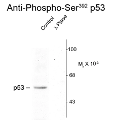
p53 (phospho Ser392) antibodyGTX23257
ApplicationsWestern Blot
TargetTP53
- SizePrice
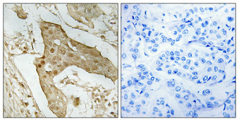
![ICC/IF analysis of HeLa cells using GTX27757 p53 antibody [BP53-12].](https://www.genetex.com/upload/website/prouct_img/normal/GTX27757/GTX27757_20191025_AP_002_194_w_23060722_732.webp)
Product group Antibodies
p53 antibody [BP53-12]GTX27757
ApplicationsFlow Cytometry, ImmunoFluorescence, ImmunoPrecipitation, Western Blot, ELISA, ImmunoCytoChemistry, ImmunoHistoChemistry, ImmunoHistoChemistry Paraffin
TargetTP53
- SizePrice
Product group Antibodies
References
p53 antibody [B20.1 (BP 53.122)]GTX28590
ApplicationsWestern Blot, ImmunoHistoChemistry, ImmunoHistoChemistry Paraffin
TargetTP53
- SizePrice
Product group Antibodies
p53 (acetyl Lys373) antibodyGTX30768
ApplicationsImmunoPrecipitation, Western Blot
TargetTP53
- SizePrice
Product group Antibodies
ApplicationsWestern Blot
TargetTP53
- SizePrice
Product group Antibodies
References
p53 antibody [DO1]GTX70214
ApplicationsFlow Cytometry, ImmunoFluorescence, ImmunoPrecipitation, Western Blot, ELISA, ImmunoCytoChemistry, ImmunoHistoChemistry, ImmunoHistoChemistry Frozen, ImmunoHistoChemistry Paraffin
TargetTP53
- SizePrice