

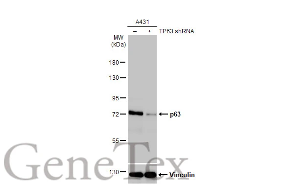
Non-transfected (–) and transfected (+) A431 whole cell extracts (30 μg) were separated by 7.5% SDS-PAGE, and the membrane was blotted with p63 antibody [HL1027] (GTX635841) diluted at 1:50000. The HRP-conjugated anti-rabbit IgG antibody (GTX213110-01) was used to detect the primary antibody, and the signal was developed with Trident ECL plus-Enhanced.
![Whole zebrafish extract (30 μg) was separated by 7.5% SDS-PAGE, and the membrane was blotted with p63 antibody [HL1027] (GTX635841) diluted at 1:1000. The HRP-conjugated anti-rabbit IgG antibody (GTX213110-01) was used to detect the primary antibody, and the signal was developed with Trident ECL plus-Enhanced.](https://www.genetex.com/upload/website/prouct_img/normal/GTX635841/GTX635841_44340_20221216_WB_Z_22122018_500.webp "Whole zebrafish extract (30 μg) was separated by 7.5% SDS-PAGE, and the membrane was blotted with p63 antibody [HL1027] (GTX635841) diluted at 1:1000. The HRP-conjugated anti-rabbit IgG antibody (GTX213110-01) was used to detect the primary antibody, and the signal was developed with Trident ECL plus-Enhanced.")
![p63 antibody [HL1027] detects p63 protein on whole mount zebrafish by immunohistochemical analysis. Sample: Paraformaldehyde-fixed 2 days-post-fertilization zebrafish embryo. Green: p63 stained by p63 antibody [HL1027] (GTX635841) diluted at 1:100. Antigen Retrieval: Tris-HCl buffer, pH 9.0, 20 min at 70oC](https://www.genetex.com/upload/website/prouct_img/normal/GTX635841/GTX635841_44340_20230203_IHC-Wm_Z_23041719_599.webp "p63 antibody [HL1027] detects p63 protein on whole mount zebrafish by immunohistochemical analysis. Sample: Paraformaldehyde-fixed 2 days-post-fertilization zebrafish embryo. Green: p63 stained by p63 antibody [HL1027] (GTX635841) diluted at 1:100. Antigen Retrieval: Tris-HCl buffer, pH 9.0, 20 min at 70oC")
![p63 antibody [HL1027] detects p63 protein at nucleus by immunofluorescent analysis. Sample: A431 cells were fixed in 4% paraformaldehyde at RT for 15 min. Green: p63 stained by p63 antibody [HL1027] (GTX635841) diluted at 1:500. Red: alpha Tubulin, a cytoskeleton marker, stained by alpha Tubulin antibody [GT114] (GTX628802) diluted at 1:1000.](https://www.genetex.com/upload/website/prouct_img/normal/GTX635841/GTX635841_T-44130_20210416_ICC_IF_w_23061202_984.webp "p63 antibody [HL1027] detects p63 protein at nucleus by immunofluorescent analysis. Sample: A431 cells were fixed in 4% paraformaldehyde at RT for 15 min. Green: p63 stained by p63 antibody [HL1027] (GTX635841) diluted at 1:500. Red: alpha Tubulin, a cytoskeleton marker, stained by alpha Tubulin antibody [GT114] (GTX628802) diluted at 1:1000.")
![p63 antibody [HL1027] detects p63 protein at nucleus by immunohistochemical analysis. Sample: Paraffin-embedded mouse testis. p63 stained by p63 antibody [HL1027] (GTX635841) diluted at 1:100. Antigen Retrieval: Citrate buffer, pH 6.0, 15 min](https://www.genetex.com/upload/website/prouct_img/normal/GTX635841/GTX635841_T-44130_20220304_IHC-P_M_w_23061202_810.webp "p63 antibody [HL1027] detects p63 protein at nucleus by immunohistochemical analysis. Sample: Paraffin-embedded mouse testis. p63 stained by p63 antibody [HL1027] (GTX635841) diluted at 1:100. Antigen Retrieval: Citrate buffer, pH 6.0, 15 min")
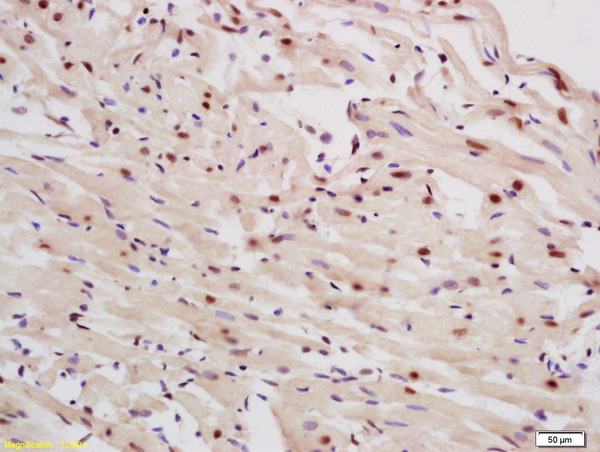
![p63 antibody [HL1027] detects p63 protein at nucleus by immunohistochemical analysis. Sample: Paraffin-embedded human placenta. p63 stained by p63 antibody [HL1027] (GTX635841) diluted at 1:100. Antigen Retrieval: Citrate buffer, pH 6.0, 15 min](https://www.genetex.com/upload/website/prouct_img/normal/GTX635841/GTX635841_45278_20240301_IHC-P_24030600_463.webp "p63 antibody [HL1027] detects p63 protein at nucleus by immunohistochemical analysis. Sample: Paraffin-embedded human placenta. p63 stained by p63 antibody [HL1027] (GTX635841) diluted at 1:100. Antigen Retrieval: Citrate buffer, pH 6.0, 15 min")
![p63 antibody [HL1027] detects p63 protein by immunohistochemical analysis. Sample: Paraffin-embedded mouse tongue. p63 stained by p63 antibody [HL1027] (GTX635841) diluted at 1:1000. Antigen Retrieval: Citrate buffer, pH 6.0, 15 min](https://www.genetex.com/upload/website/prouct_img/normal/GTX635841/GTX635841_44340_20241004_IHC-P_M_24110700_723.webp "p63 antibody [HL1027] detects p63 protein by immunohistochemical analysis. Sample: Paraffin-embedded mouse tongue. p63 stained by p63 antibody [HL1027] (GTX635841) diluted at 1:1000. Antigen Retrieval: Citrate buffer, pH 6.0, 15 min")
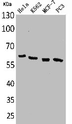
![Various whole cell extracts (30 μg) were separated by 7.5% SDS-PAGE, and the membrane was blotted with p63 antibody [HL1027] (GTX635841) diluted at 1:1000. The HRP-conjugated anti-rabbit IgG antibody (GTX213110-01) was used to detect the primary antibody. Corresponding RNA expression data for the same cell lines are based on Human Protein Atlas program.](https://www.genetex.com/upload/website/prouct_img/normal/GTX635841/GTX635841_45250_20231215_WB_TPM_watermark_24112622_516.webp "Various whole cell extracts (30 μg) were separated by 7.5% SDS-PAGE, and the membrane was blotted with p63 antibody [HL1027] (GTX635841) diluted at 1:1000. The HRP-conjugated anti-rabbit IgG antibody (GTX213110-01) was used to detect the primary antibody. Corresponding RNA expression data for the same cell lines are based on Human Protein Atlas program.")
Non-transfected (–) and transfected (+) A431 whole cell extracts (30 μg) were separated by 7.5% SDS-PAGE, and the membrane was blotted with p63 antibody [HL1027] (GTX635841) diluted at 1:50000. The HRP-conjugated anti-rabbit IgG antibody (GTX213110-01) was used to detect the primary antibody, and the signal was developed with Trident ECL plus-Enhanced.
p63 antibody [HL1027]
GTX635841
ApplicationsImmunoFluorescence, Western Blot, ImmunoCytoChemistry, ImmunoHistoChemistry, ImmunoHistoChemistry Paraffin
Product group Antibodies
ReactivityHuman, Mouse, Zebra Fish
TargetTP63
Overview
- SupplierGeneTex
- Product Namep63 antibody [HL1027]
- Delivery Days Customer9
- Application Supplier NoteWB: 1:500-1:3000. *Optimal dilutions/concentrations should be determined by the researcher.Not tested in other applications.
- ApplicationsImmunoFluorescence, Western Blot, ImmunoCytoChemistry, ImmunoHistoChemistry, ImmunoHistoChemistry Paraffin
- CertificationResearch Use Only
- ClonalityMonoclonal
- Clone IDHL1027
- Concentration0.3 mg/ml
- ConjugateUnconjugated
- Gene ID8626
- Target nameTP63
- Target descriptiontumor protein p63
- Target synonymsAIS, B(p51A), B(p51B), EEC3, KET, LMS, NBP, OFC8, RHS, SHFM4, TP53CP, TP53L, TP73L, p40, p51, p53CP, p63, p73H, p73L, tumor protein 63, amplified in squamous cell carcinoma, chronic ulcerative stomatitis protein, keratinocyte transcription factor KET, transformation-related protein 63, tumor protein p53-competing protein
- HostRabbit
- IsotypeIgG
- Protein IDQ9H3D4
- Protein NameTumor protein 63
- Scientific DescriptionThis gene encodes a member of the p53 family of transcription factors. The functional domains of p53 family proteins include an N-terminal transactivation domain, a central DNA-binding domain and an oligomerization domain. Alternative splicing of this gene and the use of alternative promoters results in multiple transcript variants encoding different isoforms that vary in their functional properties. These isoforms function during skin development and maintenance, adult stem/progenitor cell regulation, heart development and premature aging. Some isoforms have been found to protect the germline by eliminating oocytes or testicular germ cells that have suffered DNA damage. Mutations in this gene are associated with ectodermal dysplasia, and cleft lip/palate syndrome 3 (EEC3); split-hand/foot malformation 4 (SHFM4); ankyloblepharon-ectodermal defects-cleft lip/palate; ADULT syndrome (acro-dermato-ungual-lacrimal-tooth); limb-mammary syndrome; Rap-Hodgkin syndrome (RHS); and orofacial cleft 8. [provided by RefSeq, Aug 2016]
- ReactivityHuman, Mouse, Zebra Fish
- Storage Instruction-20°C or -80°C,2°C to 8°C
- UNSPSC12352203
Datasheet
Related products
Product group Antibodies

Anti-p63 [BU5]Ab00871-10.0
ApplicationsFlow Cytometry, ImmunoFluorescence
ReactivityHuman
TargetTP63
- SizePrice

Product group Antibodies
Anti-TP63 AntibodyAMAB91224
ApplicationsWestern Blot, ImmunoHistoChemistry
ReactivityHuman
TargetTP63
- SizePrice

Product group Antibodies
Anti-TP63 Antibody144-12937
ApplicationsImmunoFluorescence, Western Blot
ReactivityHuman, Mouse, Rat
TargetTP63
- SizePrice
Product group Antibodies
ApplicationsImmunoPrecipitation, Western Blot, ImmunoCytoChemistry, ImmunoHistoChemistry
TargetTP63
- SizePrice
Product group Antibodies
References
p63 Polyclonal AntibodyBS-0723R
ApplicationsImmunoFluorescence, Western Blot, ELISA, ImmunoCytoChemistry, ImmunoHistoChemistry, ImmunoHistoChemistry Frozen, ImmunoHistoChemistry Paraffin
ReactivityBovine, Canine, Equine, Guinea Pig, Human, Mouse, Porcine, Rabbit, Rat, Sheep
TargetTP63
- SizePrice
Product group Antibodies
TP63 AntibodyCSB-PA005081
ApplicationsWestern Blot, ELISA
ReactivityHuman, Mouse, Rat
TargetTP63
- SizePrice
Product group Antibodies
p63 antibody [ZR8]GTX01785
ApplicationsImmunoHistoChemistry, ImmunoHistoChemistry Paraffin
ReactivityHuman
TargetTP63
- SizePrice