

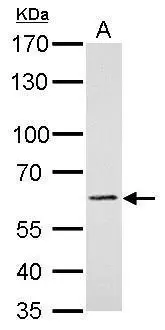
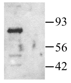
PAK1 antibody detects PAK1 protein by western blot analysis. A. 30 μg BCL-1 whole cell lysate/extract 7.5% SDS-PAGE PAK1 antibody (GTX103797) dilution: 1:1000 The HRP-conjugated anti-rabbit IgG antibody (GTX213110-01) was used to detect the primary antibody.
 diluted at 1:500. Blue: Hoechst 33343 staining.")
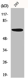
 was separated by 7.5% SDS-PAGE, and the membrane was blotted with PAK1 antibody (GTX103797) diluted at 1:1000. The HRP-conjugated anti-rabbit IgG antibody (GTX213110-01) was used to detect the primary antibody.")
 dilution: 1:1000 The HRP-conjugated anti-rabbit IgG antibody (GTX213110-01) was used to detect the primary antibody.")
 dilution: 1:500.
Antigen Retrieval: Trilogy? (EDTA based, pH 8.0) buffer, 15min")
 A: A431 B: HeLa 7.5% SDS PAGE GTX103797 diluted at 1:5000 The HRP-conjugated anti-rabbit IgG antibody (GTX213110-01) was used to detect the primary antibody.")
PAK1 antibody detects PAK1 protein by western blot analysis. A. 30 μg BCL-1 whole cell lysate/extract 7.5% SDS-PAGE PAK1 antibody (GTX103797) dilution: 1:1000 The HRP-conjugated anti-rabbit IgG antibody (GTX213110-01) was used to detect the primary antibody.
PAK1 antibody
GTX103797
ApplicationsImmunoFluorescence, Western Blot, ImmunoCytoChemistry, ImmunoHistoChemistry, ImmunoHistoChemistry Paraffin
Product group Antibodies
ReactivityHuman, Mouse, Rat
TargetPAK1
Overview
- SupplierGeneTex
- Product NamePAK1 antibody
- Delivery Days Customer9
- Application Supplier NoteWB: 1:500-1:10000. ICC/IF: 1:100-1:1000. IHC-P: 1:100-1:1000. *Optimal dilutions/concentrations should be determined by the researcher.Not tested in other applications.
- ApplicationsImmunoFluorescence, Western Blot, ImmunoCytoChemistry, ImmunoHistoChemistry, ImmunoHistoChemistry Paraffin
- CertificationResearch Use Only
- ClonalityPolyclonal
- Concentration0.48 mg/ml
- ConjugateUnconjugated
- Gene ID5058
- Target namePAK1
- Target descriptionp21 (RAC1) activated kinase 1
- Target synonymsIDDMSSD, PAKalpha, alpha-PAK, p65-PAK, serine/threonine-protein kinase PAK 1, STE20 homolog, yeast, p21 protein (Cdc42/Rac)-activated kinase 1, p21/Cdc42/Rac1-activated kinase 1 (STE20 homolog, yeast), p21/Cdc42/Rac1-activated kinase 1 (yeast Ste20-related)
- HostRabbit
- IsotypeIgG
- Protein IDQ13153
- Protein NameSerine/threonine-protein kinase PAK 1
- Scientific DescriptionPAK proteins are critical effectors that link RhoGTPases to cytoskeleton reorganization and nuclear signaling. PAK proteins, a family of serine/threonine p21-activating kinases, include PAK1, PAK2, PAK3 and PAK4. These proteins serve as targets for the small GTP binding proteins Cdc42 and Rac and have been implicated in a wide range of biological activities. PAK1 regulates cell motility and morphology. Alternativelt spliced transcript variants encoding different isoforms have been found for this gene. [provided by RefSeq]
- ReactivityHuman, Mouse, Rat
- Storage Instruction-20°C or -80°C,2°C to 8°C
- UNSPSC41116161
Datasheet
Related products
Product group Antibodies
Anti-PAK1 AntibodyA95353
ApplicationsImmunoFluorescence, Western Blot, ELISA, ImmunoHistoChemistry
ReactivityHuman, Mouse, Rat
- SizePrice
Product group Antibodies
Anti-PAK1 Antibody144-60034
ApplicationsWestern Blot, ImmunoHistoChemistry
ReactivityHuman, Rat
TargetPAK1
- SizePrice
Product group Antibodies

PAK1 AntibodyLS-C769449
ApplicationsWestern Blot, ELISA, ImmunoHistoChemistry, ImmunoHistoChemistry Paraffin
ReactivityHuman, Mouse, Rat
TargetPAK1
- SizePrice

Product group Antibodies
Anti-PAK1 Antibody Picoband(r)A00454-1-CARRIER-FREE
ApplicationsWestern Blot, ELISA
ReactivityHuman, Mouse, Rat
TargetPAK1
- SizePrice
Product group Antibodies
PAK1 AntibodyCSB-PA003703
ApplicationsImmunoFluorescence, Western Blot, ELISA, ImmunoHistoChemistry
ReactivityHuman, Mouse, Rat
TargetPAK1
- SizePrice
Product group Antibodies

Goat anti-PAK1EB07367
ApplicationsWestern Blot, ELISA
ReactivityCanine, Human, Mouse, Rat
TargetPAK1
- SizePrice
Product group Antibodies
ApplicationsWestern Blot, ImmunoHistoChemistry
ReactivityRat
TargetPAK1
- SizePrice
Product group Antibodies
ApplicationsImmunoFluorescence, Western Blot, ELISA, ImmunoCytoChemistry, ImmunoHistoChemistry, ImmunoHistoChemistry Frozen, ImmunoHistoChemistry Paraffin
ReactivityBovine, Canine, Chicken, Human, Mouse, Porcine, Rabbit, Rat
TargetPAK1
- SizePrice
Product group Antibodies
ApplicationsDot Blot, Western Blot, ELISA
ReactivityHuman
- SizePrice
Product group Antibodies
PAK1 antibodyGTX22551
ApplicationsImmunoPrecipitation, Western Blot
ReactivityHuman
TargetPAK1
- SizePrice