

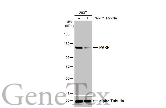
Non-transfected (–) and transfected (+) 293T whole cell extracts (30 μg) were separated by 7.5% SDS-PAGE, and the membrane was blotted with PARP antibody [HL1365] (GTX636805) diluted at 1:10000. The HRP-conjugated anti-rabbit IgG antibody (GTX213110-01) was used to detect the primary antibody.
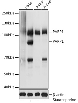
![Various whole cell extracts (30 μg) were separated by 7.5% SDS-PAGE, and the membranes were blotted with PARP antibody [HL1365] (GTX636805) diluted at 1:10000 and competitor's antibody (# Highly Cited Antibody) diluted at 1:1000. The HRP-conjugated anti-rabbit IgG antibody (GTX213110-01) was used to detect the primary antibody. *The competitor is not affiliated with GeneTex and does not endorse this product.](https://www.genetex.com/upload/website/prouct_img/normal/GTX636805/GTX636805_44683_20220812_WB_competitor_watermark_22082402_217.webp "Various whole cell extracts (30 μg) were separated by 7.5% SDS-PAGE, and the membranes were blotted with PARP antibody [HL1365] (GTX636805) diluted at 1:10000 and competitor's antibody (# Highly Cited Antibody) diluted at 1:1000. The HRP-conjugated anti-rabbit IgG antibody (GTX213110-01) was used to detect the primary antibody. *The competitor is not affiliated with GeneTex and does not endorse this product.")
![PARP antibody [HL1365] detects PARP protein at nucleus by immunofluorescent analysis. Sample: HeLa cells were fixed in 4% paraformaldehyde at RT for 15 min. Green: PARP stained by PARP antibody [HL1365] (GTX636805) diluted at 1:500. Red: alpha Tubulin, a cytoskeleton marker, stained by alpha Tubulin antibody [GT114] (GTX628802) diluted at 1:1000.](https://www.genetex.com/upload/website/prouct_img/normal/GTX636805/GTX636805_T-44599_20220325_ICC_IF_w_23061202_854.webp "PARP antibody [HL1365] detects PARP protein at nucleus by immunofluorescent analysis. Sample: HeLa cells were fixed in 4% paraformaldehyde at RT for 15 min. Green: PARP stained by PARP antibody [HL1365] (GTX636805) diluted at 1:500. Red: alpha Tubulin, a cytoskeleton marker, stained by alpha Tubulin antibody [GT114] (GTX628802) diluted at 1:1000.")
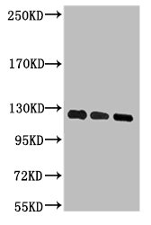
![Untreated (–) and treated (+) HCT-116 whole cell extract (30 μg) were separated by 7.5% SDS-PAGE, and the membrane was blotted with PARP antibody [HL1365] (GTX636805) diluted at 1:10000. The HRP-conjugated anti-rabbit IgG antibody (GTX213110-01) was used to detect the primary antibody.](https://www.genetex.com/upload/website/prouct_img/normal/GTX636805/GTX636805_T-44599_20220513_WB_treatment_Cisplatin_w_23061202_431.webp "Untreated (–) and treated (+) HCT-116 whole cell extract (30 μg) were separated by 7.5% SDS-PAGE, and the membrane was blotted with PARP antibody [HL1365] (GTX636805) diluted at 1:10000. The HRP-conjugated anti-rabbit IgG antibody (GTX213110-01) was used to detect the primary antibody.")
![Non-transfected (–) and transfected (+) 293T whole cell extracts (30 μg) were separated by 7.5% SDS-PAGE, and the membrane was blotted with PARP antibody [HL1365] (GTX636805) diluted at 1:100000. The HRP-conjugated anti-rabbit IgG antibody (GTX213110-01) was used to detect the primary antibody.](https://www.genetex.com/upload/website/prouct_img/normal/GTX636805/GTX636805_44683_20231006_WB_multiple_B_23102401_438.webp "Non-transfected (–) and transfected (+) 293T whole cell extracts (30 μg) were separated by 7.5% SDS-PAGE, and the membrane was blotted with PARP antibody [HL1365] (GTX636805) diluted at 1:100000. The HRP-conjugated anti-rabbit IgG antibody (GTX213110-01) was used to detect the primary antibody.")
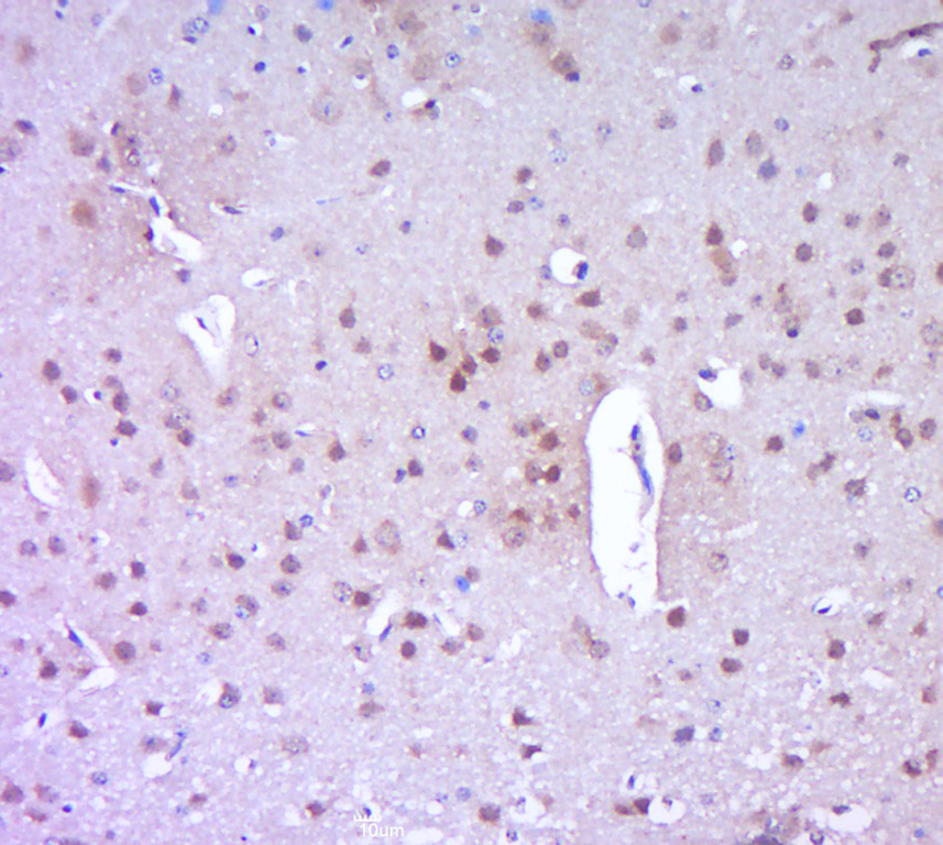
![PARP antibody [HL1365] detects PARP protein by immunohistochemical analysis. Sample: Paraffin-embedded human tonsil. PARP stained by PARP antibody [HL1365] (GTX636805) diluted at 1:400. Antigen Retrieval: Tris-EDTA buffer, pH 9.0, 15 min](https://www.genetex.com/upload/website/prouct_img/normal/GTX636805/GTX636805_45530_20250829_IHC-P_1_25090420_497.webp "PARP antibody [HL1365] detects PARP protein by immunohistochemical analysis. Sample: Paraffin-embedded human tonsil. PARP stained by PARP antibody [HL1365] (GTX636805) diluted at 1:400. Antigen Retrieval: Tris-EDTA buffer, pH 9.0, 15 min")
![PARP antibody [HL1365] detects PARP protein by immunohistochemical analysis. Sample: Paraffin-embedded human placenta. PARP stained by PARP antibody [HL1365] (GTX636805) diluted at 1:400. Antigen Retrieval: Tris-EDTA buffer, pH 9.0, 15 min](https://www.genetex.com/upload/website/prouct_img/normal/GTX636805/GTX636805_45530_20250829_IHC-P_3_25090420_782.webp "PARP antibody [HL1365] detects PARP protein by immunohistochemical analysis. Sample: Paraffin-embedded human placenta. PARP stained by PARP antibody [HL1365] (GTX636805) diluted at 1:400. Antigen Retrieval: Tris-EDTA buffer, pH 9.0, 15 min")
![PARP antibody [HL1365] detects PARP protein by immunohistochemical analysis. Sample: Paraffin-embedded human appendix. PARP stained by PARP antibody [HL1365] (GTX636805) diluted at 1:400. Antigen Retrieval: Tris-EDTA buffer, pH 9.0, 15 min](https://www.genetex.com/upload/website/prouct_img/normal/GTX636805/GTX636805_45530_20250829_IHC-P_2_25090420_657.webp "PARP antibody [HL1365] detects PARP protein by immunohistochemical analysis. Sample: Paraffin-embedded human appendix. PARP stained by PARP antibody [HL1365] (GTX636805) diluted at 1:400. Antigen Retrieval: Tris-EDTA buffer, pH 9.0, 15 min")
Non-transfected (–) and transfected (+) 293T whole cell extracts (30 μg) were separated by 7.5% SDS-PAGE, and the membrane was blotted with PARP antibody [HL1365] (GTX636805) diluted at 1:10000. The HRP-conjugated anti-rabbit IgG antibody (GTX213110-01) was used to detect the primary antibody.
PARP antibody [HL1365]
GTX636805
ApplicationsImmunoFluorescence, Western Blot, ImmunoCytoChemistry, ImmunoHistoChemistry, ImmunoHistoChemistry Paraffin
Product group Antibodies
ReactivityHuman, Mouse
TargetPARP1
Overview
- SupplierGeneTex
- Product NamePARP antibody [HL1365]
- Delivery Days Customer9
- Application Supplier NoteICC/IF: 1:100-1:1000. *Optimal dilutions/concentrations should be determined by the researcher.Not tested in other applications.
- ApplicationsImmunoFluorescence, Western Blot, ImmunoCytoChemistry, ImmunoHistoChemistry, ImmunoHistoChemistry Paraffin
- CertificationResearch Use Only
- ClonalityMonoclonal
- Clone IDHL1365
- Concentration1 mg/ml
- ConjugateUnconjugated
- Gene ID142
- Target namePARP1
- Target descriptionpoly(ADP-ribose) polymerase 1
- Target synonymsADPRT, ADPRT 1, ADPRT1, ARTD1, PARP, PARP-1, PARS, PPOL, Poly-PARP, pADPRT-1, poly [ADP-ribose] polymerase 1, ADP-ribosyltransferase (NAD+; poly (ADP-ribose) polymerase), ADP-ribosyltransferase NAD(+), ADP-ribosyltransferase diphtheria toxin-like 1, DNA ADP-ribosyltransferase PARP1, NAD(+) ADP-ribosyltransferase 1, poly (ADP-ribose) polymerase family, member 1, poly(ADP-ribose) synthetase, poly(ADP-ribosyl)transferase, poly[ADP-ribose] synthase 1, protein poly-ADP-ribosyltransferase PARP1
- HostRabbit
- IsotypeIgG
- Protein IDP09874
- Protein NamePoly [ADP-ribose] polymerase 1
- Scientific DescriptionThis gene encodes a chromatin-associated enzyme, poly(ADP-ribosyl)transferase, which modifies various nuclear proteins by poly(ADP-ribosyl)ation. The modification is dependent on DNA and is involved in the regulation of various important cellular processes such as differentiation, proliferation, and tumor transformation and also in the regulation of the molecular events involved in the recovery of cell from DNA damage. In addition, this enzyme may be the site of mutation in Fanconi anemia, and may participate in the pathophysiology of type I diabetes. [provided by RefSeq, Jul 2008]
- ReactivityHuman, Mouse
- Storage Instruction-20°C or -80°C,2°C to 8°C
- UNSPSC41116161
Datasheet
Related products

Product group Antibodies
Anti-PARP1 Antibody Picoband(r)A00122-2-CARRIER-FREE
ApplicationsFlow Cytometry, ImmunoFluorescence, Western Blot, ELISA, ImmunoCytoChemistry
ReactivityHuman
TargetPARP1
- SizePrice
Product group Antibodies
Anti-PARP1 AntibodyA11205
ApplicationsImmunoFluorescence, Western Blot, ImmunoCytoChemistry
ReactivityHuman, Mouse, Rat
- SizePrice
Product group Antibodies
Anti-PARP1 Antibody144-00942
ApplicationsImmunoFluorescence, Western Blot, ImmunoHistoChemistry
ReactivityHuman, Mouse, Rat
TargetPARP1
- SizePrice

Product group Antibodies
Anti-PARP1 AntibodyAMAB90959
ApplicationsWestern Blot, ImmunoCytoChemistry, ImmunoHistoChemistry
ReactivityHuman
TargetPARP1
- SizePrice
Product group Antibodies

PARP1 Antibody (clone 4A3)LS-C765617
ApplicationsWestern Blot
ReactivityHuman
TargetPARP1
- SizePrice
Product group Antibodies
PARP1 Monoclonal AntibodyCSB-MA000290
ApplicationsWestern Blot, ELISA
ReactivityHuman
TargetPARP1
- SizePrice
Product group Antibodies
References
PARP1 Polyclonal AntibodyBS-2138R
ApplicationsFlow Cytometry, ImmunoFluorescence, Western Blot, ELISA, ImmunoCytoChemistry, ImmunoHistoChemistry, ImmunoHistoChemistry Frozen, ImmunoHistoChemistry Paraffin
ReactivityHuman
TargetPARP1
- SizePrice
![Untreated (–) and treated (+) HCT-116 whole cell extract (30 μg) were separated by 7.5% SDS-PAGE, and the membrane was blotted with PARP antibody [HL1364] (GTX636804) diluted at 1:10000. The HRP-conjugated anti-rabbit IgG antibody (GTX213110-01) was used to detect the primary antibody.](https://www.genetex.com/upload/website/prouct_img/normal/GTX636804/GTX636804_44711_20220617_WB_treatment_Cisplatin_22062121_177.webp)
Product group Antibodies
PARP antibody [HL1364]GTX636804
ApplicationsImmunoFluorescence, Western Blot, ImmunoCytoChemistry, ImmunoHistoChemistry, ImmunoHistoChemistry Paraffin
ReactivityHuman, Mouse
TargetPARP1
- SizePrice