

PathPlus(tm) GAP43 Antibody
LS-B2157
ApplicationsWestern Blot, ELISA, ImmunoHistoChemistry, ImmunoHistoChemistry Frozen, ImmunoHistoChemistry Paraffin
Product group Antibodies
ReactivityChicken, Human, Mouse, Rabbit, Reptile, Rat
TargetGAP43
Overview
- SupplierLifeSpan BioSciences
- Product NamePathPlus(tm) GAP43 Antibody
- Delivery Days Customer23
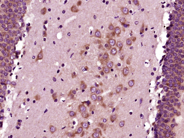
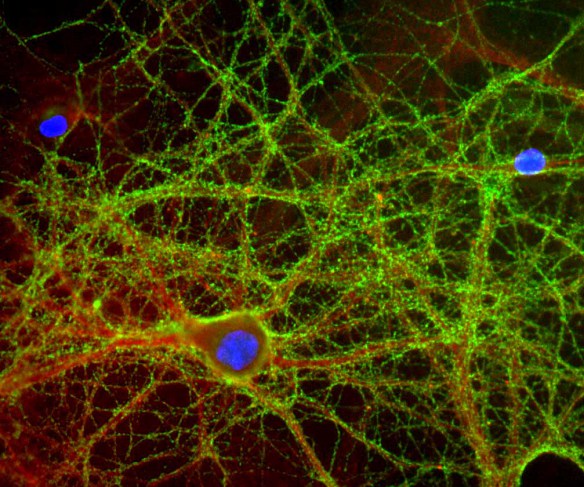
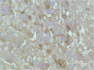
- Application Supplier NoteImmunohistochemistry: LS-B2157 was validated for use in immunohistochemistry on a panel of 21 formalin-fixed, paraffin-embedded (FFPE) human tissues after heat induced antigen retrieval in pH 6.0 citrate buffer. After incubation with the primary antibody, slides were incubated with biotinylated secondary antibody, followed by alkaline phosphatase-streptavidin and chromogen. The stained slides were evaluated by a pathologist to confirm staining specificity. The optimal working concentration for LS-B2157 was determined to be 5 ug/ml. Positive control: Cell lysates derived from mouse and rat brain.. ELISA (0.1 - 1 µg/ml), IHC, IHC-Fr (5 µg/ml), IHC-P (5 µg/ml), WB (1 µg/ml) Immunohistochemistry: LS-B2157 was validated for use in immunohistochemistry on a panel of 21 formalin-fixed, paraffin-embedded (FFPE) human tissues after heat induced antigen retrieval in pH 6.0 citrate buffer. After incubation with the primary antibody, slides were incubated with biotinylated secondary antibody, followed by alkaline phosphatase-streptavidin and chromogen. The stained slides were evaluated by a pathologist to confirm staining specificity. The optimal working concentration for LS-B2157 was determined to be 5 ug/ml. Positive control: Cell lysates derived from mouse and rat brain.
- ApplicationsWestern Blot, ELISA, ImmunoHistoChemistry, ImmunoHistoChemistry Frozen, ImmunoHistoChemistry Paraffin
- CertificationResearch Use Only
- ClonalityMonoclonal
- Concentration0.5 mg/ml
- ConjugateUnconjugated
- Estimated Purity...
- Gene ID2596
- Target nameGAP43
- Target descriptiongrowth associated protein 43
- Target synonymsB-50, GAP-43, PP46, neuromodulin, axonal membrane protein GAP-43, calmodulin-binding protein P-57, nerve growth-related peptide GAP43, neural phosphoprotein B-50, neuron growth-associated protein 43, protein F1
- HostMouse
- IsotypeIgG2a
- ReactivityChicken, Human, Mouse, Rabbit, Reptile, Rat
- Storage Instruction-20°C,2°C to 8°C
- UNSPSC12352203
Related products
Product group Antibodies
ApplicationsFlow Cytometry, ImmunoFluorescence, ELISA, ImmunoCytoChemistry, ImmunoHistoChemistry, ImmunoHistoChemistry Frozen, ImmunoHistoChemistry Paraffin
ReactivityCanine, Human, Mouse, Rat
TargetGAP43
- SizePrice
Product group Antibodies
ApplicationsWestern Blot, ELISA, ImmunoCytoChemistry, ImmunoHistoChemistry, ImmunoHistoChemistry Frozen, ImmunoHistoChemistry Paraffin
ReactivityPorcine, Rat
TargetGAP43
- SizePrice
Product group Antibodies
Anti-GAP43 AntibodyA85292
ApplicationsImmunoFluorescence, Western Blot, ImmunoCytoChemistry, ImmunoHistoChemistry
ReactivityBovine, Equine, Human, Mouse, Porcine, Rat
- SizePrice
Product group Antibodies
Anti-GAP43 Antibody144-06376
ApplicationsWestern Blot
ReactivityHuman, Mouse, Rat
TargetGAP43
- SizePrice

Product group Antibodies
Anti-GAP43 AntibodyAMAB91664
ApplicationsImmunoHistoChemistry
ReactivityHuman
TargetGAP43
- SizePrice
Product group Antibodies

GAP43 Antibody (clone 6F2)LS-C772675
ApplicationsWestern Blot, ImmunoHistoChemistry, ImmunoHistoChemistry Paraffin
ReactivityHuman, Mouse, Rat
TargetGAP43
- SizePrice

Product group Antibodies
Anti-GAP43 Antibody Picoband(r)A01868-CARRIER-FREE
ApplicationsFlow Cytometry, ImmunoFluorescence, Western Blot, ImmunoHistoChemistry
ReactivityHuman, Mouse, Rat
TargetGAP43
- SizePrice
Product group Antibodies
GAP43 Monoclonal AntibodyCSB-MA080173
ApplicationsWestern Blot, ELISA, ImmunoHistoChemistry
ReactivityHuman, Mouse, Rat
TargetGAP43
- SizePrice