

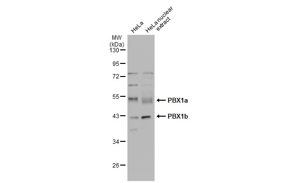
HeLa whole cell and nuclear extracts (30 μg) were separated by 10% SDS-PAGE, and the membrane was blotted with PBX1 antibody [N2C3] (GTX105260) diluted at 1:1000. The HRP-conjugated anti-rabbit IgG antibody (GTX213110-01) was used to detect the primary antibody.
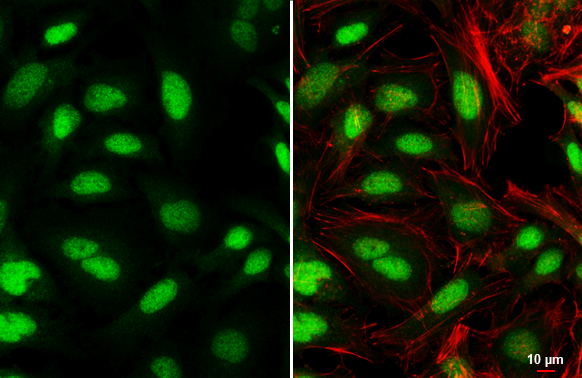
![PBX1 antibody [N2C3] detects PBX1 protein at nucleus and cytoplasm by immunofluorescent analysis. Sample: SK-N-AS cells were fixed in 4% paraformaldehyde at RT for 15 min. Green: PBX1 protein stained by PBX1 antibody [N2C3] (GTX105260) diluted at 1:500. Blue: Hoechst 33342 staining.](https://www.genetex.com/upload/website/prouct_img/normal/GTX105260/GTX105260_39974_IFA_w_23060120_912.webp "PBX1 antibody [N2C3] detects PBX1 protein at nucleus and cytoplasm by immunofluorescent analysis. Sample: SK-N-AS cells were fixed in 4% paraformaldehyde at RT for 15 min. Green: PBX1 protein stained by PBX1 antibody [N2C3] (GTX105260) diluted at 1:500. Blue: Hoechst 33342 staining.")
HeLa whole cell and nuclear extracts (30 μg) were separated by 10% SDS-PAGE, and the membrane was blotted with PBX1 antibody [N2C3] (GTX105260) diluted at 1:1000. The HRP-conjugated anti-rabbit IgG antibody (GTX213110-01) was used to detect the primary antibody.
PBX1 antibody [N2C3]
GTX105260
ApplicationsImmunoFluorescence, Western Blot, ImmunoCytoChemistry
Product group Antibodies
ReactivityHuman
TargetPBX1
Overview
- SupplierGeneTex
- Product NamePBX1 antibody [N2C3]
- Delivery Days Customer9
- Application Supplier NoteWB: 1:500-1:3000. ICC/IF: 1:100-1:1000. *Optimal dilutions/concentrations should be determined by the researcher.Not tested in other applications.
- ApplicationsImmunoFluorescence, Western Blot, ImmunoCytoChemistry
- CertificationResearch Use Only
- ClonalityPolyclonal
- Concentration1 mg/ml
- ConjugateUnconjugated
- Gene ID5087
- Target namePBX1
- Target descriptionPBX homeobox 1
- Target synonymsCAKUHED, pre-B-cell leukemia transcription factor 1, homeobox protein PBX1, homeobox protein PRL, pre-B-cell leukemia homeobox 1
- HostRabbit
- IsotypeIgG
- Protein IDP40424
- Protein NamePre-B-cell leukemia transcription factor 1
- ReactivityHuman
- Storage Instruction-20°C or -80°C,2°C to 8°C
- UNSPSC41116161
Datasheet
Related products
Product group Antibodies
PBX1 AntibodyCSB-PA017505LA01HU
ApplicationsImmunoFluorescence, Western Blot, ELISA, ImmunoHistoChemistry
ReactivityHuman, Rat
TargetPBX1
- SizePrice
Product group Antibodies
Anti-PBX1 Antibody144-66432
ApplicationsWestern Blot
ReactivityHuman, Mouse
TargetPBX1
- SizePrice
Product group Antibodies

Goat anti-PBX1EB10052
ApplicationsWestern Blot, ELISA
ReactivityCanine, Human, Mouse, Rat
TargetPBX1
- SizePrice

Product group Antibodies
Anti-PBX1 AntibodyHPA003505
ApplicationsWestern Blot, ImmunoCytoChemistry, ImmunoHistoChemistry
ReactivityHuman, Mouse, Rat
TargetPBX1
- SizePrice
Product group Antibodies

PBX1 AntibodyLS-C330800
ApplicationsWestern Blot
ReactivityHuman, Rat
TargetPBX1
- SizePrice
Product group Antibodies
Pbx1 Polyclonal AntibodyCAC11241
ApplicationsImmunoFluorescence, Western Blot, ELISA, ImmunoHistoChemistry
ReactivityRat
TargetPBX1
- SizePrice
Product group Antibodies
PBX1 antibodyGTX113242
ApplicationsImmunoFluorescence, ImmunoPrecipitation, Western Blot, ImmunoCytoChemistry
ReactivityHuman
TargetPBX1
- SizePrice
Product group Antibodies
PBX1 antibody, InternalGTX88088
ApplicationsWestern Blot
ReactivityHuman, Mouse, Rat
TargetPBX1
- SizePrice
Product group Antibodies
PBX1 Polyclonal AntibodyBS-12296R
ApplicationsImmunoFluorescence, Western Blot, ELISA, ImmunoCytoChemistry, ImmunoHistoChemistry, ImmunoHistoChemistry Frozen, ImmunoHistoChemistry Paraffin
ReactivityBovine, Canine, Chicken, Equine, Human, Mouse, Rabbit, Rat, Zebra Fish
TargetPBX1
- SizePrice