

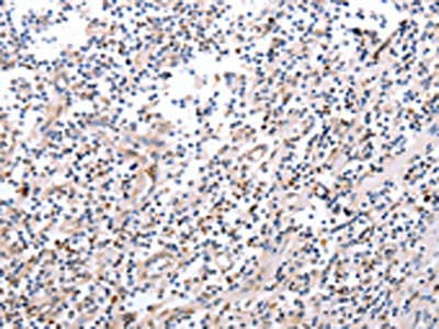
The image on the left is immunohistochemistry of paraffin-embedded Human tonsil tissue using CSB-PA469157(PDCD10 Antibody) at dilution 1/20, on the right is treated with fusion protein. (Original magnification: x200)
 at dilution 1/20, on the right is treated with fusion protein. (Original magnification: x200)")
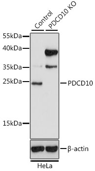
 at dilution 1/200, Secondary antibody: Goat anti rabbit IgG at 1/8000 dilution, Exposure time: 10 seconds")
The image on the left is immunohistochemistry of paraffin-embedded Human tonsil tissue using CSB-PA469157(PDCD10 Antibody) at dilution 1/20, on the right is treated with fusion protein. (Original magnification: x200)
PDCD10 Antibody
CSB-PA469157
ApplicationsWestern Blot, ELISA, ImmunoHistoChemistry
Product group Antibodies
ReactivityHuman, Mouse, Rat
TargetPDCD10
Overview
- SupplierCusabio
- Product NamePDCD10 Antibody
- Delivery Days Customer20
- ApplicationsWestern Blot, ELISA, ImmunoHistoChemistry
- CertificationResearch Use Only
- ClonalityPolyclonal
- ConjugateUnconjugated
- Gene ID11235
- Target namePDCD10
- Target descriptionprogrammed cell death 10
- Target synonymsCCM3, TFAR15, programmed cell death protein 10, TF-1 cell apoptosis-related protein 15, apoptosis-related protein 15, cerebral cavernous malformations 3 protein
- HostRabbit
- IsotypeIgG
- Protein IDQ9BUL8
- Protein NameProgrammed cell death protein 10
- Scientific DescriptionThis gene encodes an evolutionarily conserved protein associated with cell apoptosis. The protein interacts with the serine/threonine protein kinase MST4 to modulate the extracellular signal-regulated kinase (ERK) pathway. It also interacts with and is phosphoryated by serine/threonine kinase 25, and is thought to function in a signaling pathway essential for vascular developent. Mutations in this gene are one cause of cerebral cavernous malformations, which are vascular malformations that cause seizures and cerebral hemorrhages. Multiple alternatively spliced variants, encoding the same protein, have been identified.
- ReactivityHuman, Mouse, Rat
- Storage Instruction-20°C or -80°C
- UNSPSC41116161
Related products
Product group Antibodies
Anti-PDCD10 Antibody144-64794
ApplicationsWestern Blot, ImmunoHistoChemistry
ReactivityHuman, Mouse, Rat
TargetPDCD10
- SizePrice
Product group Antibodies
PDCD10 Polyclonal AntibodyBS-55165R
ApplicationsImmunoFluorescence, Western Blot
ReactivityHuman, Mouse
TargetPDCD10
- SizePrice
Product group Antibodies
PDCD10 antibodyGTX35229
ApplicationsWestern Blot
ReactivityHuman, Mouse
TargetPDCD10
- SizePrice

Product group Antibodies
Anti-PDCD10 AntibodyHPA027095
ApplicationsImmunoHistoChemistry
ReactivityHuman
TargetPDCD10
- SizePrice
Product group Antibodies

PDCD10 AntibodyLS-C332244
ApplicationsWestern Blot, ImmunoHistoChemistry
ReactivityHuman, Rat
TargetPDCD10
- SizePrice
Product group Antibodies
Anti-PDCD10 AntibodyA30639
ApplicationsWestern Blot, ImmunoHistoChemistry
ReactivityHuman, Mouse, Rat
- SizePrice

Product group Antibodies
Anti-PDCD10 Antibody Picoband(r)A01879-1-CARRIER-FREE
ApplicationsWestern Blot, ELISA
ReactivityHuman, Mouse, Rat
TargetPDCD10
- SizePrice