

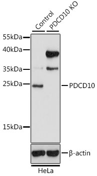
Lane 1: Hela cell lysates; Lane 2: PDCD10 knockout (KO) HeLa cell lysates probed with PDCD10 Polyclonal Antibody, Unconjugated (bs-55165R) at 1:1000 dilution and 4˚C overnight incubation. Followed by conjugated secondary antibody incubation at 1:20000 for 60 min at 37˚C.
 at 1:1000 dilution and 4˚C overnight incubation. Followed by conjugated secondary antibody incubation at 1:20000 for 60 min at 37˚C.")
Lane 1: Hela cell lysates; Lane 2: PDCD10 knockout (KO) HeLa cell lysates probed with PDCD10 Polyclonal Antibody, Unconjugated (bs-55165R) at 1:1000 dilution and 4˚C overnight incubation. Followed by conjugated secondary antibody incubation at 1:20000 for 60 min at 37˚C.
PDCD10 Polyclonal Antibody
BS-55165R
ApplicationsImmunoFluorescence, Western Blot
Product group Antibodies
ReactivityHuman, Mouse
TargetPDCD10
Overview
- SupplierBioss
- Product NamePDCD10 Polyclonal Antibody
- Delivery Days Customer16
- ApplicationsImmunoFluorescence, Western Blot
- Applications SupplierWB(1:300-5000), IF()
- CertificationResearch Use Only
- ClonalityPolyclonal
- Concentration1 ug/ul
- ConjugateUnconjugated
- Gene ID11235
- Target namePDCD10
- Target descriptionprogrammed cell death 10
- Target synonymsCCM3, TFAR15, programmed cell death protein 10, TF-1 cell apoptosis-related protein 15, apoptosis-related protein 15, cerebral cavernous malformations 3 protein
- HostRabbit
- IsotypeIgG
- Protein IDQ9BUL8
- Protein NameProgrammed cell death protein 10
- ReactivityHuman, Mouse
- Storage Instruction-20°C
- UNSPSC41116161
Datasheet
Related products
Product group Antibodies
PDCD10 AntibodyCSB-PA469157
ApplicationsWestern Blot, ELISA, ImmunoHistoChemistry
ReactivityHuman, Mouse, Rat
TargetPDCD10
- SizePrice

Product group Antibodies
Anti-PDCD10 Antibody Picoband(r)A01879-1-CARRIER-FREE
ApplicationsWestern Blot, ELISA
ReactivityHuman, Mouse, Rat
TargetPDCD10
- SizePrice
Product group Antibodies
Anti-PDCD10 AntibodyA30639
ApplicationsWestern Blot, ImmunoHistoChemistry
ReactivityHuman, Mouse, Rat
- SizePrice

Product group Antibodies
Anti-PDCD10 AntibodyHPA027095
ApplicationsImmunoHistoChemistry
ReactivityHuman
TargetPDCD10
- SizePrice
Product group Antibodies

PDCD10 AntibodyLS-C332244
ApplicationsWestern Blot, ImmunoHistoChemistry
ReactivityHuman, Rat
TargetPDCD10
- SizePrice
Product group Antibodies
Anti-PDCD10 Antibody144-64794
ApplicationsWestern Blot, ImmunoHistoChemistry
ReactivityHuman, Mouse, Rat
TargetPDCD10
- SizePrice