

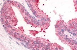
IHC-P analysis of human prostate tissue using GTX85838 PDE8A antibody. Antigen retrieval : Heat-induced antigen retrieval
IHC-P analysis of human prostate tissue using GTX85838 PDE8A antibody. Antigen retrieval : Heat-induced antigen retrieval
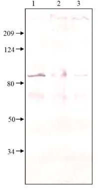
PDE8A antibody
GTX85838
ApplicationsImmunoHistoChemistry, ImmunoHistoChemistry Paraffin
Product group Antibodies
ReactivityCanine, Equine, Human, Porcine, Rabbit
TargetPDE8A
Overview
- SupplierGeneTex
- Product NamePDE8A antibody
- Delivery Days Customer9
- Application Supplier NoteIHC-P: 10 microg/ml. *Optimal dilutions/concentrations should be determined by the researcher.Not tested in other applications.
- ApplicationsImmunoHistoChemistry, ImmunoHistoChemistry Paraffin
- CertificationResearch Use Only
- ClonalityPolyclonal
- Concentration1 mg/ml
- ConjugateUnconjugated
- Gene ID5151
- Target namePDE8A
- Target descriptionphosphodiesterase 8A
- Target synonymsHsT19550, high affinity cAMP-specific and IBMX-insensitive 3',5'-cyclic phosphodiesterase 8A, cAMP-specific cyclic nucleotide phosphodiesterase 8A
- HostRabbit
- IsotypeIgG
- Protein IDO60658
- Protein NameHigh affinity cAMP-specific and IBMX-insensitive 3',5'-cyclic phosphodiesterase 8A
- Scientific DescriptionThe protein encoded by this gene belongs to the cyclic nucleotide phosphodiesterase (PDE) family, and PDE8 subfamily. This PDE hydrolyzes the second messenger, cAMP, which is a regulator and mediator of a number of cellular responses to extracellular signals. Thus, by regulating the cellular concentration of cAMP, this protein plays a key role in many important physiological processes. Alternatively spliced transcript variants encoding different isoforms have been found for this gene.[provided by RefSeq, Jul 2011]
- ReactivityCanine, Equine, Human, Porcine, Rabbit
- Storage Instruction-20°C or -80°C,2°C to 8°C
- UNSPSC41116161
Datasheet
Related products
Product group Antibodies
Anti-PDE8A AntibodyA80955
ApplicationsWestern Blot
ReactivityMouse, Rat
- SizePrice

Product group Antibodies
Anti-PDE8A Antibody Picoband(r)A05304-1-CARRIER-FREE
ApplicationsFlow Cytometry, Western Blot, ELISA
ReactivityHuman
TargetPDE8A
- SizePrice
Product group Antibodies
Anti-Mouse/Rat PDE8A Antibody144-12187
ApplicationsWestern Blot
ReactivityHuman, Mouse, Rat
TargetPDE8A
- SizePrice
Product group Antibodies

PDE8A AntibodyLS-C747356
ApplicationsWestern Blot
ReactivityHuman, Mouse, Rat
TargetPDE8A
- SizePrice
Product group Antibodies
PDE8A AntibodyCSB-PA525315LA01HU
ApplicationsImmunoFluorescence, ELISA, ImmunoHistoChemistry
ReactivityHuman
TargetPDE8A
- SizePrice
Product group Antibodies
PDE8A antibodyGTX14618
ApplicationsImmunoFluorescence, ImmunoPrecipitation, Western Blot, ELISA, ImmunoCytoChemistry, ImmunoHistoChemistry
ReactivityBovine, Human, Monkey, Mouse, Rat
TargetPDE8A
- SizePrice
Product group Antibodies
PDE8A antibodyGTX14619
ApplicationsImmunoFluorescence, ImmunoPrecipitation, Western Blot, ELISA, ImmunoCytoChemistry, ImmunoHistoChemistry
ReactivityBovine, Human, Monkey, Mouse, Rat
TargetPDE8A
- SizePrice
Product group Antibodies
PDE8A antibodyGTX14620
ApplicationsImmunoFluorescence, ImmunoPrecipitation, Western Blot, ELISA, ImmunoCytoChemistry, ImmunoHistoChemistry
ReactivityBovine, Human, Monkey, Mouse, Rat
TargetPDE8A
- SizePrice
Product group Antibodies
PDE8A antibodyGTX103504
ApplicationsImmunoFluorescence, ImmunoCytoChemistry
ReactivityHuman
TargetPDE8A
- SizePrice