

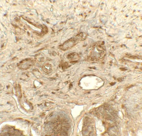
IHC-P analysis of rat small intestine tissue using GTX31623 PDI antibody. Working concentration : 5 μg/ml

 1 and (B) 2 μg/ml")
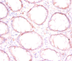
IHC-P analysis of rat small intestine tissue using GTX31623 PDI antibody. Working concentration : 5 μg/ml
PDI antibody
GTX31623
ApplicationsWestern Blot, ELISA, ImmunoHistoChemistry, ImmunoHistoChemistry Paraffin
Product group Antibodies
ReactivityHuman, Mouse, Rat
TargetP4HB
Overview
- SupplierGeneTex
- Product NamePDI antibody
- Delivery Days Customer9
- Application Supplier NoteWB: 1 - 2 microg/mL. IHC-P: 5 microg/mL. *Optimal dilutions/concentrations should be determined by the researcher.Not tested in other applications.
- ApplicationsWestern Blot, ELISA, ImmunoHistoChemistry, ImmunoHistoChemistry Paraffin
- CertificationResearch Use Only
- ClonalityPolyclonal
- Concentration1 mg/ml
- ConjugateUnconjugated
- Gene ID5034
- Target nameP4HB
- Target descriptionprolyl 4-hydroxylase subunit beta
- Target synonymsCLCRP1, DSI, ERBA2L, GIT, P4Hbeta, PDI, PDIA1, PHDB, PO4DB, PO4HB, PROHB, protein disulfide-isomerase, cellular thyroid hormone-binding protein, collagen prolyl 4-hydroxylase beta, glutathione-insulin transhydrogenase, p55, procollagen-proline, 2-oxoglutarate 4-dioxygenase (proline 4-hydroxylase), beta polypeptide, prolyl 4-hydroxylase, beta polypeptide, protein disulfide isomerase family A, member 1, protein disulfide isomerase-associated 1, protein disulfide isomerase/oxidoreductase, protocollagen hydroxylase, testicular secretory protein Li 32, thyroid hormone-binding protein p55
- HostRabbit
- IsotypeIgG
- Protein IDP07237
- Protein NameProtein disulfide-isomerase
- Scientific DescriptionThis gene encodes the beta subunit of prolyl 4-hydroxylase, a highly abundant multifunctional enzyme that belongs to the protein disulfide isomerase family. When present as a tetramer consisting of two alpha and two beta subunits, this enzyme is involved in hydroxylation of prolyl residues in preprocollagen. This enzyme is also a disulfide isomerase containing two thioredoxin domains that catalyze the formation, breakage and rearrangement of disulfide bonds. Other known functions include its ability to act as a chaperone that inhibits aggregation of misfolded proteins in a concentration-dependent manner, its ability to bind thyroid hormone, its role in both the influx and efflux of S-nitrosothiol-bound nitric oxide, and its function as a subunit of the microsomal triglyceride transfer protein complex. [provided by RefSeq, Jul 2008]
- ReactivityHuman, Mouse, Rat
- Storage Instruction-20°C or -80°C,2°C to 8°C
- UNSPSC41116161
Datasheet
Related products
Product group Antibodies
P4HB AntibodyCSB-PA00254A0RB
ApplicationsWestern Blot, ELISA, ImmunoHistoChemistry
ReactivityHuman, Mouse
TargetP4HB
- SizePrice
Product group Antibodies
P4Hb Polyclonal AntibodyCAC07340
ApplicationsWestern Blot, ELISA, ImmunoHistoChemistry
ReactivityMouse
TargetP4HB
- SizePrice
Product group Antibodies
Anti-P4HB AntibodyA28660
ApplicationsImmunoFluorescence, ImmunoPrecipitation, Western Blot, ImmunoHistoChemistry, Other Application
ReactivityHuman, Mouse, Rat
- SizePrice

Product group Antibodies
Anti-P4HB Antibody Picoband(r)A02335-1-CARRIER-FREE
ApplicationsFlow Cytometry, ImmunoFluorescence, Western Blot, ELISA, ImmunoCytoChemistry, ImmunoHistoChemistry
ReactivityHuman, Monkey, Mouse, Rat
TargetP4HB
- SizePrice
Product group Antibodies

ApplicationsELISA
ReactivityHuman
TargetP4HB
- SizePrice

Product group Antibodies
Anti-P4HB AntibodyHPA018884
ApplicationsWestern Blot, ImmunoCytoChemistry, ImmunoHistoChemistry
ReactivityHuman, Mouse, Rat
TargetP4HB
- SizePrice
Product group Antibodies
P4HB Polyclonal AntibodyBS-1476R
ApplicationsImmunoFluorescence, Western Blot, ELISA, ImmunoCytoChemistry, ImmunoHistoChemistry, ImmunoHistoChemistry Frozen, ImmunoHistoChemistry Paraffin
ReactivityBovine, Chicken, Equine, Human, Mouse, Rabbit, Rat
TargetP4HB
- SizePrice
![PDI antibody [N1N3] detects PDI protein at endoplasmic reticulum by immunofluorescent analysis. Sample: HeLa cells were fixed in 4% paraformaldehyde at RT for 15 min. Green: PDI protein stained by PDI antibody [N1N3] (GTX101468) diluted at 1:100. Red: phalloidin, a cytoskeleton marker, stained by phalloidin (invitrogen, A12380) diluted at 1:200. Blue: Hoechst 33342 staining.](https://www.genetex.com/upload/website/prouct_img/normal/GTX101468/GTX101468_39721_20150410_IFA_w_23060100_648.webp)
Product group Antibodies
PDI antibody [N1N3]GTX101468
ApplicationsElectron Microscopy, ImmunoFluorescence, Western Blot, ImmunoCytoChemistry, ImmunoHistoChemistry, ImmunoHistoChemistry Paraffin
ReactivityHuman, Mouse, Rat
TargetP4HB
- SizePrice
Product group Antibodies
PDI antibodyGTX55742
ApplicationsImmunoFluorescence, ImmunoPrecipitation, Western Blot, ImmunoCytoChemistry
ReactivityHuman, Mouse
TargetP4HB
- SizePrice