

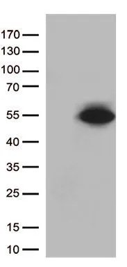
WB analysis of HEK293T cells transfected with PDX1 plasmid (Right) or empty vector (Left) for 48 hrs using GTX83918 PDX1 antibody [5D5]. Loading : 5 ug per lane
![WB analysis of various cell lines using GTX83918 PDX1 antibody [5D5]. Loading : 35 ug per lane](https://www.genetex.com/upload/website/prouct_img/normal/GTX83918/GTX83918_4002_WB_w_23061420_602.webp "WB analysis of various cell lines using GTX83918 PDX1 antibody [5D5]. Loading : 35 ug per lane")
WB analysis of HEK293T cells transfected with PDX1 plasmid (Right) or empty vector (Left) for 48 hrs using GTX83918 PDX1 antibody [5D5]. Loading : 5 ug per lane
PDX1 antibody [5D5]
GTX83918
ApplicationsWestern Blot
Product group Antibodies
ReactivityHuman
TargetPDX1
Overview
- SupplierGeneTex
- Product NamePDX1 antibody [5D5]
- Delivery Days Customer9
- Application Supplier NoteWB: 1:5000-1:10000. *Optimal dilutions/concentrations should be determined by the researcher.Not tested in other applications.
- ApplicationsWestern Blot
- CertificationResearch Use Only
- ClonalityMonoclonal
- Clone ID5D5
- Concentration1 mg/ml
- ConjugateUnconjugated
- Gene ID3651
- Target namePDX1
- Target descriptionpancreatic and duodenal homeobox 1
- Target synonymsGSF, IDX-1, IPF1, IUF1, MODY4, PAGEN1, PDX-1, STF-1, pancreas/duodenum homeobox protein 1, IPF-1, IUF-1, glucose-sensitive factor, insulin promoter factor 1, homeodomain transcription factor, insulin upstream factor 1, islet/duodenum homeobox-1, pancreatic-duodenal homeobox factor 1, somatostatin transcription factor 1, somatostatin-transactivating factor 1
- HostMouse
- IsotypeIgG1
- Protein IDP52945
- Protein NamePancreas/duodenum homeobox protein 1
- Scientific DescriptionActivates insulin, somatostatin, glucokinase, islet amyloid polypeptide and glucose transporter type 2 gene transcription. Particularly involved in glucose-dependent regulation of insulin gene transcription. Binds preferentially the DNA motif 5-[CT]TAAT[TG]-3. During development, specifies the early pancreatic epithelium, permitting its proliferation, branching and subsequent differentiation. At adult stage, required for maintaining the hormone-producing phenotype of the beta-cell.
- ReactivityHuman
- Storage Instruction-20°C or -80°C,2°C to 8°C
- UNSPSC41116161
Datasheet
Related products
Product group Antibodies
Anti-PDX1 AntibodyA97975
ApplicationsWestern Blot, ELISA
ReactivityHuman, Mouse, Rat
- SizePrice
Product group Antibodies
Anti-PDX1 Antibody144-10173
ApplicationsWestern Blot
ReactivityHuman, Mouse, Rat
TargetPDX1
- SizePrice

Product group Antibodies
PDX1 Recombinant AntibodyBSM-54153R
ApplicationsImmunoFluorescence, Western Blot, ImmunoCytoChemistry, ImmunoHistoChemistry, ImmunoHistoChemistry Frozen, ImmunoHistoChemistry Paraffin
ReactivityHuman
TargetPDX1
- SizePrice

Product group Antibodies
Anti-PDX1 Antibody Picoband(r)A00491-3-CARRIER-FREE
ApplicationsFlow Cytometry, Western Blot, ELISA
ReactivityHuman
TargetPDX1
- SizePrice
Product group Antibodies
PDX1 AntibodyCSB-PA011230
ApplicationsWestern Blot, ELISA
ReactivityHuman, Mouse, Rat
TargetPDX1
- SizePrice
Product group Antibodies

PDX1 AntibodyLS-C405773
ApplicationsWestern Blot, ELISA, ImmunoHistoChemistry
ReactivityHuman, Mouse, Rat
TargetPDX1
- SizePrice
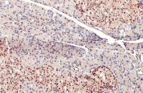
![IHC-P analysis of human pancreas carcinoma tissue using GTX83919 PDX1 antibody [2A12]. Antigen retrieval : Heat-induced epitope retrieval by 10mM citrate buffer, pH6.0, 100oC for 10min.](https://www.genetex.com/upload/website/prouct_img/normal/GTX83919/GTX83919_2019_IHC-P_w_23061420_853.webp)
Product group Antibodies
PDX1 antibody [2A12]GTX83919
ApplicationsImmunoFluorescence, Western Blot, ImmunoCytoChemistry, ImmunoHistoChemistry, ImmunoHistoChemistry Paraffin
ReactivityHuman, Mouse
TargetPDX1
- SizePrice
Product group Antibodies
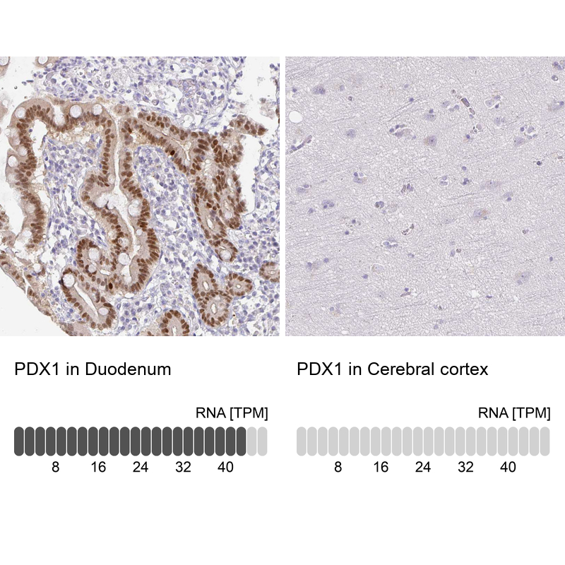
Anti-PDX1 AntibodyHPA059146
ApplicationsImmunoCytoChemistry, ImmunoHistoChemistry
ReactivityHuman
TargetPDX1
- SizePrice
Product group Antibodies
PDX1 antibodyGTX130034
ApplicationsWestern Blot, ImmunoHistoChemistry, ImmunoHistoChemistry Paraffin
ReactivityHuman, Mouse, Rat
TargetPDX1
- SizePrice