

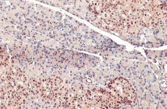
PDX1 antibody detects PDX1 protein at nucleus by immunohistochemical analysis. Sample: Paraffin-embedded rat pancreas. PDX1 stained by PDX1 antibody (GTX130034) diluted at 1:500. Antigen Retrieval: Citrate buffer, pH 6.0, 15 min
 were separated by 12% SDS-PAGE, and the membrane was blotted with PDX1 antibody (GTX130034) diluted at 1:1000. The HRP-conjugated anti-rabbit IgG antibody (GTX213110-01) was used to detect the primary antibody.")
 diluted at 1:500. Antigen Retrieval: Citrate buffer, pH 6.0, 15 min")
PDX1 antibody detects PDX1 protein at nucleus by immunohistochemical analysis. Sample: Paraffin-embedded rat pancreas. PDX1 stained by PDX1 antibody (GTX130034) diluted at 1:500. Antigen Retrieval: Citrate buffer, pH 6.0, 15 min
PDX1 antibody
GTX130034
ApplicationsWestern Blot, ImmunoHistoChemistry, ImmunoHistoChemistry Paraffin
Product group Antibodies
ReactivityHuman, Mouse, Rat
TargetPDX1
Overview
- SupplierGeneTex
- Product NamePDX1 antibody
- Delivery Days Customer9
- Application Supplier NoteWB: 1:500-1:3000. IHC-P: 1:100-1:1000. *Optimal dilutions/concentrations should be determined by the researcher.Not tested in other applications.
- ApplicationsWestern Blot, ImmunoHistoChemistry, ImmunoHistoChemistry Paraffin
- CertificationResearch Use Only
- ClonalityPolyclonal
- Concentration1.29 mg/ml
- ConjugateUnconjugated
- Gene ID3651
- Target namePDX1
- Target descriptionpancreatic and duodenal homeobox 1
- Target synonymsGSF, IDX-1, IPF1, IUF1, MODY4, PAGEN1, PDX-1, STF-1, pancreas/duodenum homeobox protein 1, IPF-1, IUF-1, glucose-sensitive factor, insulin promoter factor 1, homeodomain transcription factor, insulin upstream factor 1, islet/duodenum homeobox-1, pancreatic-duodenal homeobox factor 1, somatostatin transcription factor 1, somatostatin-transactivating factor 1
- HostRabbit
- IsotypeIgG
- Protein IDP52945
- Protein NamePancreas/duodenum homeobox protein 1
- Scientific DescriptionThe protein encoded by this gene is a transcriptional activator of several genes, including insulin, somatostatin, glucokinase, islet amyloid polypeptide, and glucose transporter type 2. The encoded nuclear protein is involved in the early development of the pancreas and plays a major role in glucose-dependent regulation of insulin gene expression. Defects in this gene are a cause of pancreatic agenesis, which can lead to early-onset insulin-dependent diabetes mellitus (NIDDM), as well as maturity onset diabetes of the young type 4 (MODY4). [provided by RefSeq]
- ReactivityHuman, Mouse, Rat
- Storage Instruction-20°C or -80°C,2°C to 8°C
- UNSPSC41116161
Datasheet
Related products
Product group Antibodies
Anti-PDX1 AntibodyA97975
ApplicationsWestern Blot, ELISA
ReactivityHuman, Mouse, Rat
- SizePrice
Product group Antibodies
Anti-PDX1 Antibody144-10173
ApplicationsWestern Blot
ReactivityHuman, Mouse, Rat
TargetPDX1
- SizePrice

Product group Antibodies
PDX1 Recombinant AntibodyBSM-54153R
ApplicationsImmunoFluorescence, Western Blot, ImmunoCytoChemistry, ImmunoHistoChemistry, ImmunoHistoChemistry Frozen, ImmunoHistoChemistry Paraffin
ReactivityHuman
TargetPDX1
- SizePrice

Product group Antibodies
Anti-PDX1 Antibody Picoband(r)A00491-3-CARRIER-FREE
ApplicationsFlow Cytometry, Western Blot, ELISA
ReactivityHuman
TargetPDX1
- SizePrice
Product group Antibodies
PDX1 AntibodyCSB-PA011230
ApplicationsWestern Blot, ELISA
ReactivityHuman, Mouse, Rat
TargetPDX1
- SizePrice
Product group Antibodies

PDX1 AntibodyLS-C405773
ApplicationsWestern Blot, ELISA, ImmunoHistoChemistry
ReactivityHuman, Mouse, Rat
TargetPDX1
- SizePrice
![WB analysis of HEK293T cells transfected with PDX1 plasmid (Right) or empty vector (Left) for 48 hrs using GTX83918 PDX1 antibody [5D5]. Loading : 5 ug per lane](https://www.genetex.com/upload/website/prouct_img/normal/GTX83918/GTX83918_4001_WB_w_23061420_212.webp)
Product group Antibodies
PDX1 antibody [5D5]GTX83918
ApplicationsWestern Blot
ReactivityHuman
TargetPDX1
- SizePrice
![IHC-P analysis of human pancreas carcinoma tissue using GTX83919 PDX1 antibody [2A12]. Antigen retrieval : Heat-induced epitope retrieval by 10mM citrate buffer, pH6.0, 100oC for 10min.](https://www.genetex.com/upload/website/prouct_img/normal/GTX83919/GTX83919_2019_IHC-P_w_23061420_853.webp)
Product group Antibodies
PDX1 antibody [2A12]GTX83919
ApplicationsImmunoFluorescence, Western Blot, ImmunoCytoChemistry, ImmunoHistoChemistry, ImmunoHistoChemistry Paraffin
ReactivityHuman, Mouse
TargetPDX1
- SizePrice
Product group Antibodies
Anti-PDX1 AntibodyHPA059146
ApplicationsImmunoCytoChemistry, ImmunoHistoChemistry
ReactivityHuman
TargetPDX1
- SizePrice