

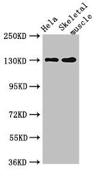
Various whole cell extracts (30 μg) were separated by 5% SDS-PAGE, and the membrane was blotted with PER3 antibody [GT1327] (GTX03239) diluted at 1:500. The HRP-conjugated anti-rabbit IgG antibody (GTX213110-01) was used to detect the primary antibody, and the signal was developed with Trident ECL plus-Enhanced.
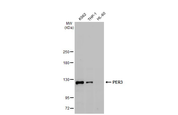
![WB analysis of various samples using GTX03239 PER3 antibody [GT1327]. Dilution : 1:1000 Loading : 25μg per lane](https://www.genetex.com/upload/website/prouct_img/normal/GTX03239/GTX03239_18_WB_w_23053123_765.webp "WB analysis of various samples using GTX03239 PER3 antibody [GT1327]. Dilution : 1:1000 Loading : 25μg per lane")
![Various whole cell extracts (30 μg) were separated by 5% SDS-PAGE, and the membrane was blotted with PER3 antibody [GT1327] (GTX03239) diluted at 1:500. The HRP-conjugated anti-rabbit IgG antibody (GTX213110-01) was used to detect the primary antibody.](https://www.genetex.com/upload/website/prouct_img/normal/GTX03239/GTX03239_4000001893_20210716_WB_3_w_23053123_951.webp "Various whole cell extracts (30 μg) were separated by 5% SDS-PAGE, and the membrane was blotted with PER3 antibody [GT1327] (GTX03239) diluted at 1:500. The HRP-conjugated anti-rabbit IgG antibody (GTX213110-01) was used to detect the primary antibody.")
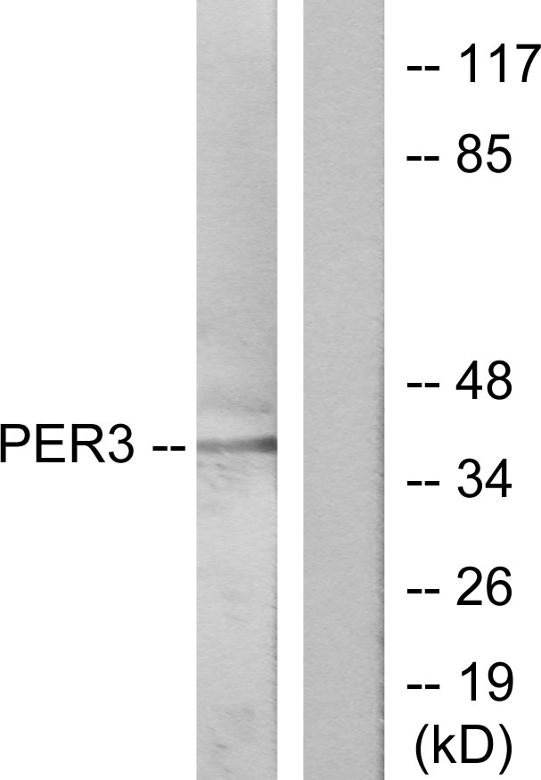
![Untreated (–) and treated (+) 293T whole cell extracts (30 μg) were separated by 5% SDS-PAGE, and the membrane was blotted with PER3 antibody [GT1327] (GTX03239) diluted at 1:500. The HRP-conjugated anti-rabbit IgG antibody (GTX213110-01) was used to detect the primary antibody, and the signal was developed with Trident ECL plus-Enhanced.](https://www.genetex.com/upload/website/prouct_img/normal/GTX03239/GTX03239_4000001893_20210716_WB_treatment_Serum_w_23053123_161.webp "Untreated (–) and treated (+) 293T whole cell extracts (30 μg) were separated by 5% SDS-PAGE, and the membrane was blotted with PER3 antibody [GT1327] (GTX03239) diluted at 1:500. The HRP-conjugated anti-rabbit IgG antibody (GTX213110-01) was used to detect the primary antibody, and the signal was developed with Trident ECL plus-Enhanced.")
![ICC/IF analysis of NIH-3T3 cells using GTX03239 PER3 antibody [GT1327]. Blue : DAPI for nuclear staining Dilution : 1:100](https://www.genetex.com/upload/website/prouct_img/normal/GTX03239/GTX03239_20210615_ICCIF_13_w_23053123_300.webp "ICC/IF analysis of NIH-3T3 cells using GTX03239 PER3 antibody [GT1327]. Blue : DAPI for nuclear staining Dilution : 1:100")
Various whole cell extracts (30 μg) were separated by 5% SDS-PAGE, and the membrane was blotted with PER3 antibody [GT1327] (GTX03239) diluted at 1:500. The HRP-conjugated anti-rabbit IgG antibody (GTX213110-01) was used to detect the primary antibody, and the signal was developed with Trident ECL plus-Enhanced.
PER3 antibody [GT1327]
GTX03239
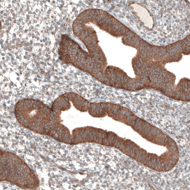
ApplicationsImmunoFluorescence, Western Blot, ImmunoCytoChemistry
Product group Antibodies
ReactivityHuman, Mouse, Rat
TargetPER3
Overview
- SupplierGeneTex
- Product NamePER3 antibody [GT1327]
- Delivery Days Customer9
- Application Supplier NoteWB: 1:500 - 1:2000. ICC/IF: 1:50 - 1:200. *Optimal dilutions/concentrations should be determined by the researcher.Not tested in other applications.
- ApplicationsImmunoFluorescence, Western Blot, ImmunoCytoChemistry
- CertificationResearch Use Only
- ClonalityMonoclonal
- Clone IDGT1327
- Concentration2.44 mg/ml
- ConjugateUnconjugated
- Gene ID8863
- Target namePER3
- Target descriptionperiod circadian regulator 3
- Target synonymsFASPS3, GIG13, period circadian protein homolog 3, cell growth-inhibiting gene 13 protein, circadian clock protein PERIOD 3, period circadian clock 3, period circadian protein 3
- HostRabbit
- IsotypeIgG
- Protein IDP56645
- Protein NamePeriod circadian protein homolog 3
- Scientific DescriptionThis gene is a member of the Period family of genes and is expressed in a circadian pattern in the suprachiasmatic nucleus, the primary circadian pacemaker in the mammalian brain. Genes in this family encode components of the circadian rhythms of locomotor activity, metabolism, and behavior. This gene is upregulated by CLOCK/ARNTL heterodimers but then represses this upregulation in a feedback loop using PER/CRY heterodimers to interact with CLOCK/ARNTL. Polymorphisms in this gene have been linked to sleep disorders. Multiple transcript variants encoding different isoforms have been found for this gene. [provided by RefSeq, Jan 2014]
- ReactivityHuman, Mouse, Rat
- Storage Instruction-20°C or -80°C,2°C to 8°C
- UNSPSC41116161
Datasheet
Related products
Product group Antibodies
PER3 AntibodyCSB-PA017788LA01HU
ApplicationsImmunoFluorescence, Western Blot, ELISA, ImmunoHistoChemistry
ReactivityHuman, Mouse
TargetPER3
- SizePrice
Product group Antibodies
Per3 Polyclonal AntibodyCAC08008
ApplicationsImmunoFluorescence, Western Blot, ELISA, ImmunoHistoChemistry
ReactivityMouse
TargetPER3
- SizePrice

Product group Antibodies
Anti-PER3 Antibody Picoband(r)A01835-1-CARRIER-FREE
ApplicationsWestern Blot, ELISA
ReactivityHuman, Mouse, Rat
TargetPER3
- SizePrice
Product group Antibodies
Anti-PER3 AntibodyA101339
ApplicationsWestern Blot, ELISA
ReactivityHuman
- SizePrice
Product group Antibodies

PER3 AntibodyLS-C830770
ApplicationsELISA, ImmunoHistoChemistry
ReactivityHuman, Mouse, Rat
TargetPER3
- SizePrice
Product group Antibodies
Anti-PER3 AntibodyHPA019530
ApplicationsImmunoHistoChemistry
ReactivityHuman
TargetPER3
- SizePrice
Product group Antibodies
PER-3 Polyclonal AntibodyBS-4498R
ApplicationsImmunoFluorescence, Western Blot, ELISA, ImmunoCytoChemistry, ImmunoHistoChemistry, ImmunoHistoChemistry Frozen, ImmunoHistoChemistry Paraffin
ReactivityHuman, Mouse, Rat
TargetPER3
- SizePrice

Product group Antibodies
PER3 antibodyGTX87243
ApplicationsWestern Blot
ReactivityHuman
TargetPER3
- SizePrice