

PGK1 antibody
70R-1199
Product group Antibodies
Overview
- SupplierBiosynth
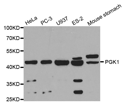
- Product NamePGK1 antibody
- Delivery Days Customer11
- CertificationResearch Use Only
- UNSPSC41116161
Related products
Product group Antibodies
Anti-PGK1 AntibodyA29944
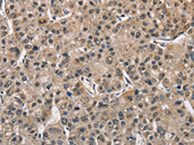
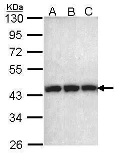
ApplicationsImmunoFluorescence, Western Blot, ImmunoHistoChemistry
ReactivityHuman, Mouse, Rat
- SizePrice
Product group Antibodies
Anti-PGK1 Antibody144-60560
ApplicationsWestern Blot, ImmunoHistoChemistry
ReactivityHuman, Mouse, Rat
TargetPGK1
- SizePrice
Product group Antibodies
PGK1 Recombinant AntibodyBSM-61216R
ApplicationsFlow Cytometry, ImmunoFluorescence, Western Blot, ImmunoCytoChemistry
TargetPGK1
- SizePrice
Product group Antibodies

Goat anti-PGK1EB12390
ApplicationsWestern Blot, ELISA
ReactivityHuman
TargetPGK1
- SizePrice
Product group Antibodies
PGK1 AntibodyCSB-PA547426
ApplicationsWestern Blot, ELISA, ImmunoHistoChemistry
ReactivityHuman, Mouse, Rat
TargetPGK1
- SizePrice
Product group Antibodies
Pgk1 Polyclonal AntibodyCAC07097
ApplicationsWestern Blot, ELISA
TargetPGK1
- SizePrice
Product group Antibodies

PGK1 / Phosphoglycerate Kinase AntibodyLS-C401396
ApplicationsWestern Blot, ELISA, ImmunoHistoChemistry
ReactivityHuman, Mouse, Rat
TargetPGK1
- SizePrice
Product group Antibodies
PGK1 antibody [N1C1]GTX101405
ApplicationsWestern Blot, ImmunoHistoChemistry, ImmunoHistoChemistry Paraffin
ReactivityHuman, Mouse, Rat
TargetPGK1
- SizePrice

Product group Antibodies
Anti-PGK1 AntibodyHPA045385
ApplicationsImmunoHistoChemistry
ReactivityHuman
TargetPGK1
- SizePrice