

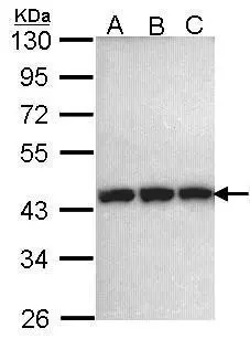
Sample (30 ug of whole cell lysate) A: 293T B: A431 (GTX27909) C: H1299 10% SDS PAGE GTX101405 diluted at 1:1000
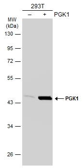
![Non-transfected (–) and transfected (+) 293T whole cell extracts (30 μg) were separated by 10% SDS-PAGE, and the membrane was blotted with PGK1 antibody [N1C1] (GTX101405) diluted at 1:1000. The HRP-conjugated anti-rabbit IgG antibody (GTX213110-01) was used to detect the primary antibody.](https://www.genetex.com/upload/website/prouct_img/normal/GTX101405/GTX101405_40150_20181221_WB_shRNA_watermark_w_23060100_178.webp "Non-transfected (–) and transfected (+) 293T whole cell extracts (30 μg) were separated by 10% SDS-PAGE, and the membrane was blotted with PGK1 antibody [N1C1] (GTX101405) diluted at 1:1000. The HRP-conjugated anti-rabbit IgG antibody (GTX213110-01) was used to detect the primary antibody.")
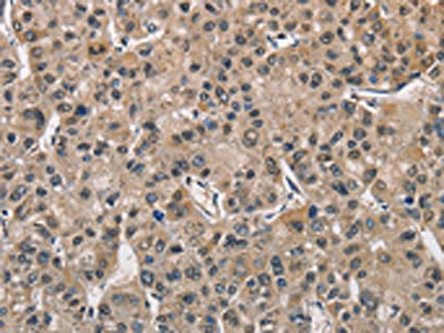
![PGK1 antibody [N1C1] detects PGK1 protein at cytoplasm by immunohistochemical analysis. Sample: Paraffin-embedded mouse testis. PGK1 stained by PGK1 antibody [N1C1] (GTX101405) diluted at 1:500. Antigen Retrieval: Citrate buffer, pH 6.0, 15 min](https://www.genetex.com/upload/website/prouct_img/normal/GTX101405/GTX101405_40436_20180622_IHC-P_M_w_23060100_817.webp "PGK1 antibody [N1C1] detects PGK1 protein at cytoplasm by immunohistochemical analysis. Sample: Paraffin-embedded mouse testis. PGK1 stained by PGK1 antibody [N1C1] (GTX101405) diluted at 1:500. Antigen Retrieval: Citrate buffer, pH 6.0, 15 min")
 A: Mouse brain 10% SDS PAGE GTX101405 diluted at 1:1000")
![PGK1 antibody [N1C1] detects PGK1 protein at cytoplasm by immunohistochemical analysis. Sample: Paraffin-embedded rat testis. PGK1 stained by PGK1 antibody [N1C1] (GTX101405) diluted at 1:500. Antigen Retrieval: Citrate buffer, pH 6.0, 15 min](https://www.genetex.com/upload/website/prouct_img/normal/GTX101405/GTX101405_40436_20180622_IHC-P_R_w_23060100_273.webp "PGK1 antibody [N1C1] detects PGK1 protein at cytoplasm by immunohistochemical analysis. Sample: Paraffin-embedded rat testis. PGK1 stained by PGK1 antibody [N1C1] (GTX101405) diluted at 1:500. Antigen Retrieval: Citrate buffer, pH 6.0, 15 min")
Sample (30 ug of whole cell lysate) A: 293T B: A431 (GTX27909) C: H1299 10% SDS PAGE GTX101405 diluted at 1:1000
PGK1 antibody [N1C1]
GTX101405
ApplicationsWestern Blot, ImmunoHistoChemistry, ImmunoHistoChemistry Paraffin
Product group Antibodies
ReactivityHuman, Mouse, Rat
TargetPGK1
Overview
- SupplierGeneTex
- Product NamePGK1 antibody [N1C1]
- Delivery Days Customer9
- Application Supplier NoteWB: 1:500-1:3000. IHC-P: 1:100-1:1000. *Optimal dilutions/concentrations should be determined by the researcher.Not tested in other applications.
- ApplicationsWestern Blot, ImmunoHistoChemistry, ImmunoHistoChemistry Paraffin
- CertificationResearch Use Only
- ClonalityPolyclonal
- Concentration1 mg/ml
- ConjugateUnconjugated
- Gene ID5230
- Target namePGK1
- Target descriptionphosphoglycerate kinase 1
- Target synonymsHEL-S-68p, MIG10, PGKA, phosphoglycerate kinase 1, PRP 2, cell migration-inducing gene 10 protein, epididymis secretory sperm binding protein Li 68p, primer recognition protein 2
- HostRabbit
- IsotypeIgG
- Protein IDP00558
- Protein NamePhosphoglycerate kinase 1
- Scientific DescriptionThe protein encoded by this gene is a glycolytic enzyme that catalyzes the conversion of 1,3-diphosphoglycerate to 3-phosphoglycerate. The encoded protein may also act as a cofactor for polymerase alpha. This gene lies on the X-chromosome, while a related pseudogene also has been found on the X-chromosome and another on chromosome 19. [provided by RefSeq]
- ReactivityHuman, Mouse, Rat
- Storage Instruction-20°C or -80°C,2°C to 8°C
- UNSPSC41116161
Datasheet
Related products
Product group Antibodies
Anti-PGK1 AntibodyA29944
ApplicationsImmunoFluorescence, Western Blot, ImmunoHistoChemistry
ReactivityHuman, Mouse, Rat
- SizePrice
Product group Antibodies
Anti-PGK1 Antibody144-60560
ApplicationsWestern Blot, ImmunoHistoChemistry
ReactivityHuman, Mouse, Rat
TargetPGK1
- SizePrice
Product group Antibodies
PGK1 Recombinant AntibodyBSM-61216R
ApplicationsFlow Cytometry, ImmunoFluorescence, Western Blot, ImmunoCytoChemistry
TargetPGK1
- SizePrice
Product group Antibodies

Goat anti-PGK1EB12390
ApplicationsWestern Blot, ELISA
ReactivityHuman
TargetPGK1
- SizePrice
Product group Antibodies
PGK1 AntibodyCSB-PA547426
ApplicationsWestern Blot, ELISA, ImmunoHistoChemistry
ReactivityHuman, Mouse, Rat
TargetPGK1
- SizePrice
Product group Antibodies
Pgk1 Polyclonal AntibodyCAC07097
ApplicationsWestern Blot, ELISA
TargetPGK1
- SizePrice
Product group Antibodies

PGK1 / Phosphoglycerate Kinase AntibodyLS-C401396
ApplicationsWestern Blot, ELISA, ImmunoHistoChemistry
ReactivityHuman, Mouse, Rat
TargetPGK1
- SizePrice

Product group Antibodies
Anti-PGK1 AntibodyHPA045385
ApplicationsImmunoHistoChemistry
ReactivityHuman
TargetPGK1
- SizePrice
Product group Antibodies
PGK1 antibodyGTX107614
ApplicationsImmunoFluorescence, ImmunoPrecipitation, Western Blot, ELISA, ImmunoCytoChemistry, Other Application
ReactivityHuman, Mouse, Rat, Yeast
TargetPGK1
- SizePrice