

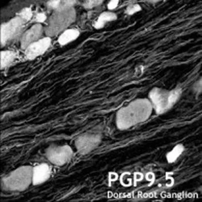
IHC-Fr analysis of rat dorsal root ganglion using GTX10404 PGP9.5 antibody.

IHC-Fr analysis of rat dorsal root ganglion using GTX10404 PGP9.5 antibody.
PGP9.5 antibody
GTX10404
ApplicationsWestern Blot, ImmunoHistoChemistry, ImmunoHistoChemistry Frozen
Product group Antibodies
ReactivityHuman, Mouse, Porcine, Rat
TargetUCHL1
Overview
- SupplierGeneTex
- Product NamePGP9.5 antibody
- Delivery Days Customer9
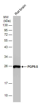
- Application Supplier NoteWB: 1:1000. IHC-Fr: 1:100-1:500. *Optimal dilutions/concentrations should be determined by the researcher.Not tested in other applications.
- ApplicationsWestern Blot, ImmunoHistoChemistry, ImmunoHistoChemistry Frozen
- CertificationResearch Use Only
- ClonalityPolyclonal
- ConjugateUnconjugated
- Gene ID7345
- Target nameUCHL1
- Target descriptionubiquitin C-terminal hydrolase L1
- Target synonymsHEL-117, HEL-S-53, NDGOA, PARK5, PGP 9.5, PGP9.5, PGP95, SPG79, SPG79A, UCHL-1, Uch-L1, ubiquitin carboxyl-terminal hydrolase isozyme L1, epididymis luminal protein 117, epididymis secretory protein Li 53, neuron cytoplasmic protein 9.5, ubiquitin carboxyl-terminal esterase L1 (ubiquitin thiolesterase), ubiquitin thioesterase L1, ubiquitin thiolesterase
- HostRabbit
- IsotypeIgG
- Protein IDP09936
- Protein NameUbiquitin carboxyl-terminal hydrolase isozyme L1
- Scientific DescriptionThe protein encoded by this gene belongs to the peptidase C12 family. This enzyme is a thiol protease that hydrolyzes a peptide bond at the C-terminal glycine of ubiquitin. This gene is specifically expressed in the neurons and in cells of the diffuse neuroendocrine system. Mutations in this gene may be associated with Parkinson disease.[provided by RefSeq, Sep 2009]
- ReactivityHuman, Mouse, Porcine, Rat
- Storage Instruction-20°C or -80°C,2°C to 8°C
- UNSPSC12352203
Datasheet
Related products
Product group Antibodies
Anti-UCH-L1 Antibody130-10634
ApplicationsELISA
ReactivityHuman
TargetUCHL1
- SizePrice
![IHC-P analysis of human cerebellum tissue section using GTX02737 PGP9.5 antibody [UCHL1/4556R].](https://www.genetex.com/upload/website/prouct_img/normal/GTX02737/GTX02737_20210319_IHC-P_1_w_23053122_123.webp)
Product group Antibodies
PGP9.5 antibody [UCHL1/4556R]GTX02737
ApplicationsWestern Blot, ImmunoHistoChemistry, ImmunoHistoChemistry Paraffin
ReactivityHuman, Rat
TargetUCHL1
- SizePrice
![IHC-P analysis of human testis tissue using GTX04438 PGP9.5 antibody [MSVA-905R] HistoMAX?. A strong PGP9.5 immunostaining is seen in spermatogonia. PGP9.5 staining decreases sharply in spermatocytes where it is only faint. A moderate PGP9.5 staining is seen in Leydig cells while Sertoli cells remain PGP9.5 negative.](https://www.genetex.com/upload/website/prouct_img/normal/GTX04438/GTX04438_20230728_IHC-P_97_23072722_457.webp)
Product group Antibodies
ApplicationsImmunoHistoChemistry, ImmunoHistoChemistry Paraffin
ReactivityHuman
TargetUCHL1
- SizePrice
Product group Antibodies
PGP9.5 antibodyGTX101093
ApplicationsWestern Blot, ImmunoHistoChemistry, ImmunoHistoChemistry Frozen, ImmunoHistoChemistry Paraffin
ReactivityHuman, Mouse, Rat
TargetUCHL1
- SizePrice
Product group Antibodies
References
PGP9.5 antibodyGTX109637
ApplicationsImmunoFluorescence, Western Blot, ImmunoCytoChemistry, ImmunoHistoChemistry, ImmunoHistoChemistry Frozen, ImmunoHistoChemistry Paraffin
ReactivityHuman, Mouse, Rat
TargetUCHL1
- SizePrice
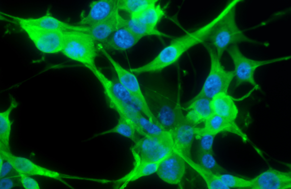
![PGP9.5 antibody detects PGP9.5 protein at cytoplasm by immunofluorescent analysis. Sample: DIV9 rat E18 primary cortical neurons were fixed in 4% paraformaldehyde at RT for 15 min. Green: PGP9.5 protein stained by PGP9.5 antibody (GTX109646) diluted at 1:500. Red: beta Tubulin 3/ Tuj1, stained by beta Tubulin 3/ Tuj1 antibody [GT11710] (GTX631836) diluted at 1:500. Blue: Fluoroshield with DAPI (GTX30920).](https://www.genetex.com/upload/website/prouct_img/normal/GTX109646/GTX109646_39959_20170503_IFA_R_w_23060500_628.webp)
Product group Antibodies
PGP9.5 antibodyGTX109646
ApplicationsImmunoFluorescence, Western Blot, ImmunoCytoChemistry, ImmunoHistoChemistry, ImmunoHistoChemistry Frozen, ImmunoHistoChemistry Paraffin
ReactivityHuman, Mouse, Rat
TargetUCHL1
- SizePrice
Product group Antibodies
PGP9.5 antibody [3D9]GTX57556
ApplicationsImmunoFluorescence, Western Blot, ImmunoCytoChemistry, ImmunoHistoChemistry, ImmunoHistoChemistry Paraffin
ReactivityHuman, Mouse
TargetUCHL1
- SizePrice
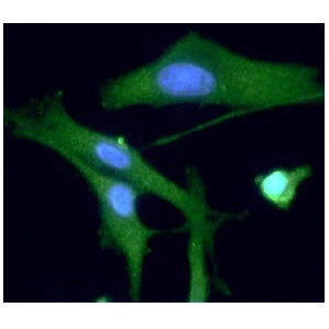
![PGP9.5 antibody [GT448] detects PGP9.5 protein by immunofluorescent analysis. Sample: DIV9 rat E18 primary hippocampal neuron cells were fixed in 4% paraformaldehyde at RT for 15 min. Green: PGP9.5 stained by PGP9.5 antibody [GT448] (GTX634797) diluted at 1:500. Red: NeuN, stained by NeuN antibody (GTX132974) diluted at 1:1000. Blue: Fluoroshield with DAPI (GTX30920).](https://www.genetex.com/upload/website/prouct_img/normal/GTX634797/GTX634797_43339_20190306_ICC_IF_R_w_23061202_510.webp)
Product group Antibodies
PGP9.5 antibody [GT448]GTX634797
ApplicationsImmunoFluorescence, Western Blot, ImmunoCytoChemistry, ImmunoHistoChemistry, ImmunoHistoChemistry Paraffin
ReactivityHuman, Mouse, Rat
TargetUCHL1
- SizePrice
Product group Antibodies
Anti-UCHL1Y058669
ApplicationsWestern Blot, ELISA, ImmunoHistoChemistry
ReactivityHuman
- SizePrice