

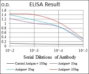
ELISA analysis of antigen using GTX60752 PHC1 antibody [1F3F3].
Black : Control antigen 100ng
Purple : Antigen 10ng
Blue : Antigen 50ng
Red : Antigen 100ng
![FACS analysis of HEK293 cells using GTX60752 PHC1 antibody [1F3F3]. Green : PHC1 Red : negative control](https://www.genetex.com/upload/website/prouct_img/normal/GTX60752/GTX60752_20170912_FACS_w_23061123_660.webp "FACS analysis of HEK293 cells using GTX60752 PHC1 antibody [1F3F3]. Green : PHC1 Red : negative control")
![WB analysis of HEK293 (1) and PHC1 (AA: 758-1004)-hIgGFc transfected HEK293 (2) cell lysate using GTX60752 PHC1 antibody [1F3F3].](https://www.genetex.com/upload/website/prouct_img/normal/GTX60752/GTX60752_20170912_WB_w_23061123_585.webp "WB analysis of HEK293 (1) and PHC1 (AA: 758-1004)-hIgGFc transfected HEK293 (2) cell lysate using GTX60752 PHC1 antibody [1F3F3].")
ELISA analysis of antigen using GTX60752 PHC1 antibody [1F3F3].
Black : Control antigen 100ng
Purple : Antigen 10ng
Blue : Antigen 50ng
Red : Antigen 100ng
PHC1 antibody [1F3F3]
GTX60752
ApplicationsFlow Cytometry, Western Blot, ELISA
Product group Antibodies
ReactivityHuman
TargetPHC1
Overview
- SupplierGeneTex
- Product NamePHC1 antibody [1F3F3]
- Delivery Days Customer9
- Application Supplier NoteWB: 1/500 - 1/2000. FCM: 1/200 - 1/400. ELISA: 1/10000. *Optimal dilutions/concentrations should be determined by the researcher.Not tested in other applications.
- ApplicationsFlow Cytometry, Western Blot, ELISA
- CertificationResearch Use Only
- ClonalityMonoclonal
- Clone ID1F3F3
- Concentration1 mg/ml
- ConjugateUnconjugated
- Gene ID1911
- Target namePHC1
- Target descriptionpolyhomeotic homolog 1
- Target synonymsEDR1, HPH1, MCPH11, RAE28, polyhomeotic-like protein 1, early development regulator 1 (homolog of polyhomeotic 1), early development regulatory protein 1, polyhomeotic-like 1
- HostMouse
- IsotypeIgG1
- Protein IDP78364
- Protein NamePolyhomeotic-like protein 1
- Scientific DescriptionThis gene is a homolog of the Drosophila polyhomeotic gene, which is a member of the Polycomb group of genes. The gene product is a component of a multimeric protein complex that contains EDR2 and the vertebrate Polycomb protein BMH1. The gene product, the EDR2 protein, and the Drosophila polyhomeotic protein share 2 highly conserved domains, named homology domains I and II. These domains are involved in protein-protein interactions and may mediate heterodimerization of the protein encoded by this gene and the EDR2 protein. [provided by RefSeq, Jul 2008]
- ReactivityHuman
- Storage Instruction-20°C or -80°C,2°C to 8°C
- UNSPSC41116161
References
- Neuronal necrosis is regulated by a conserved chromatin-modifying cascade. Liu K et al., 2014 Sep 23, Proc Natl Acad Sci U S ARead this paper
Datasheet
Related products
Product group Antibodies
PHC1 AntibodyCSB-PA017891HA01HU
ApplicationsELISA, ImmunoHistoChemistry
ReactivityHuman
TargetPHC1
- SizePrice

Product group Antibodies
Anti-PHC1 Antibody Picoband(r)A06437-3-CARRIER-FREE
ApplicationsImmunoFluorescence, Western Blot, ELISA, ImmunoCytoChemistry
ReactivityHuman
TargetPHC1
- SizePrice
Product group Antibodies
Anti-PHC1 AntibodyA31087
ApplicationsWestern Blot
ReactivityHuman, Mouse, Rat
- SizePrice

Product group Antibodies
Anti-PHC1 AntibodyHPA006973
ApplicationsImmunoCytoChemistry, ImmunoHistoChemistry
ReactivityHuman
TargetPHC1
- SizePrice
Product group Antibodies

PHC1 / EDR1 AntibodyLS-C335607
ApplicationsWestern Blot, ImmunoHistoChemistry
ReactivityHuman, Mouse, Rat
TargetPHC1
- SizePrice
Product group Antibodies
PHC1 antibodyGTX32784
ApplicationsWestern Blot, ImmunoHistoChemistry, ImmunoHistoChemistry Paraffin
ReactivityHuman, Mouse, Rat
TargetPHC1
- SizePrice
Product group Antibodies
Anti-PHC1 Antibody144-05843
ApplicationsWestern Blot, ImmunoHistoChemistry
ReactivityHuman, Mouse, Rat
TargetPHC1
- SizePrice
Product group Antibodies
PHC1 Polyclonal AntibodyBS-12302R
ApplicationsImmunoFluorescence, ELISA, ImmunoCytoChemistry, ImmunoHistoChemistry, ImmunoHistoChemistry Frozen, ImmunoHistoChemistry Paraffin
ReactivityBovine, Canine, Human, Mouse, Porcine, Rabbit, Rat, Sheep
TargetPHC1
- SizePrice