

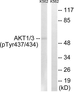
Western blot analysis of extracts from K562 cells, treated with insulin (0.01U/ml, 15mins), using AKT1/3 (Phospho-Tyr437/434) antibody. The lane on the right is treated with the synthesized peptide.
 antibody. The picture on the right is treated with the synthesized peptide.")
Western blot analysis of extracts from K562 cells, treated with insulin (0.01U/ml, 15mins), using AKT1/3 (Phospho-Tyr437/434) antibody. The lane on the right is treated with the synthesized peptide.
Phospho-AKT1/AKT3 (Tyr437/434) Antibody
CSB-PA038073
ApplicationsWestern Blot, ELISA, ImmunoHistoChemistry
Product group Antibodies
ReactivityHuman, Mouse, Rat
TargetAKT1
Overview
- SupplierCusabio
- Product NamePhospho-AKT1/AKT3 (Tyr437/434) Antibody
- Delivery Days Customer20
- ApplicationsWestern Blot, ELISA, ImmunoHistoChemistry
- CertificationResearch Use Only
- ClonalityPolyclonal
- ConjugateUnconjugated
- Gene ID207
- Target nameAKT1
- Target descriptionAKT serine/threonine kinase 1
- Target synonymsAKT, PKB, PKB-ALPHA, PRKBA, RAC, RAC-ALPHA, RAC-alpha serine/threonine-protein kinase, AKT1m, PKB alpha, RAC-PK-alpha, protein kinase B alpha, proto-oncogene c-Akt, rac protein kinase alpha, serine-threonine protein kinase, v-akt murine thymoma viral oncogene homolog 1, v-akt murine thymoma viral oncogene-like protein 1
- HostRabbit
- IsotypeIgG
- Protein IDP31749
- Protein NameRAC-alpha serine/threonine-protein kinase
- Scientific DescriptionAKT1 is one of 3 closely related serine/threonine-protein kinases (AKT1, AKT2 and AKT3) called the AKT kinase, and which regulate many processes including metabolism, proliferation, cell survival, growth and angiogenesis. This is mediated through serine and/or threonine phosphorylation of a range of downstream substrates. Over 100 substrate candidates have been reported so far, but for most of them, no isoform specificity has been reported. AKT is responsible of the regulation of glucose uptake by mediating insulin-induced translocation of the SLC2A4/GLUT4 glucose transporter to the cell surface. Phosphorylation of PTPN1 at Ser-50 negatively modulates its phosphatase activity preventing dephosphorylation of the insulin receptor and the attenuation of insulin signaling.
- ReactivityHuman, Mouse, Rat
- Storage Instruction-20°C or -80°C
- UNSPSC41116161
Related products
Product group Antibodies

Anti-pAKT [AbAb-pAKT]AB04102-10.0
ApplicationsImmunoPrecipitation, Western Blot, ELISA
ReactivityHuman, Mouse
TargetAKT1
- SizePrice
Product group Antibodies
Anti-AKT1 Antibody144-11016
ApplicationsImmunoFluorescence, Western Blot, ImmunoHistoChemistry
ReactivityHuman, Mouse, Rat
TargetAKT1
- SizePrice

Product group Antibodies
Anti-AKT1,2,3 Antibody Picoband(r)A00024-2-CARRIER-FREE
ApplicationsFlow Cytometry, ImmunoFluorescence, Western Blot, ELISA, ImmunoCytoChemistry
ReactivityHuman, Mouse, Rat
TargetAKT1
- SizePrice
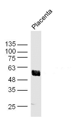
![Akt1 antibody immunoprecipitates Akt1 protein in IP experiments. IP samples: 30 μg whole cell extract of Akt1-transfected 293T cells. A. 30 μg whole cell extract of Akt1-protein expressing 293T cell B. Control with 3 μg of preimmune Rabbit IgG C. Immunoprecipitation of Akt1 protein by 3 μg Akt1 antibody (GTX110613) 10 % SDS-PAGE The immunoprecipitated Akt1 protein was detected by Akt1 antibody (GTX110613) diluted at 1:5000. [EasyBlot anti-rabbit IgG (GTX221666-01) was used as a secondary reagent]](https://www.genetex.com/upload/website/prouct_img/normal/GTX110613/GTX110613_40051_IP_w_23060500_283.webp)
Product group Antibodies
References
AKT1 antibodyGTX110613
ApplicationsImmunoPrecipitation, Western Blot, ImmunoHistoChemistry, ImmunoHistoChemistry Paraffin
ReactivityHuman, Mouse
TargetAKT1
- SizePrice
Product group Antibodies
Anti-AKT1Y058027
ApplicationsImmunoFluorescence, Western Blot, ELISA, ImmunoCytoChemistry
ReactivityHuman, Mouse
- SizePrice
Product group Antibodies
ReactivityHuman
TargetAKT1
- SizePrice
Product group Antibodies
Akt1 Polyclonal AntibodyCAC07008
ApplicationsImmunoFluorescence, ImmunoPrecipitation, Western Blot, ELISA, ImmunoHistoChemistry
TargetAKT1
- SizePrice
Product group Antibodies
AKT1 Polyclonal AntibodyBS-0115M
ApplicationsImmunoFluorescence, Western Blot, ELISA, ImmunoCytoChemistry, ImmunoHistoChemistry, ImmunoHistoChemistry Frozen, ImmunoHistoChemistry Paraffin
ReactivityBovine, Canine, Chicken, Human, Mouse, Porcine, Rabbit, Rat, Sheep
TargetAKT1
- SizePrice
Product group Antibodies
Anti-AKT1 AntibodyA285951
ApplicationsWestern Blot, ELISA
ReactivityHuman, Mouse
- SizePrice