

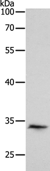
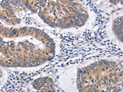
Pirh2 Antibody
ABX011352
ApplicationsFlow Cytometry, ImmunoFluorescence, Western Blot, ELISA, ImmunoCytoChemistry, ImmunoHistoChemistry
Product group Antibodies
Overview
- SupplierAbbexa
- Product NamePirh2 Antibody
- Delivery Days Customer12
- ApplicationsFlow Cytometry, ImmunoFluorescence, Western Blot, ELISA, ImmunoCytoChemistry, ImmunoHistoChemistry
- CertificationResearch Use Only
- ClonalityMonoclonal
- ConjugateUnconjugated
- HostMouse
- UNSPSC12352203
Related products
Product group Antibodies
Anti-RCHY1 AntibodyA37826
ApplicationsWestern Blot, ImmunoHistoChemistry
ReactivityHuman
- SizePrice

Product group Antibodies
Anti-Pirh2/RCHY1 Antibody Picoband(r)A04533-3-CARRIER-FREE
ApplicationsFlow Cytometry, Western Blot, ELISA
ReactivityHuman, Mouse, Rat
TargetRCHY1
- SizePrice
Product group Antibodies

RCHY1 / PIRH2 AntibodyLS-C831745
ApplicationsWestern Blot, ELISA, ImmunoHistoChemistry
ReactivityHuman, Mouse
TargetRCHY1
- SizePrice
Product group Antibodies
RCHY1 Recombinant Antibody, AbBy Fluor-647 ConjugatedBSM-62425R-BF647
ApplicationsWestern Blot
ReactivityHuman, Mouse
TargetRCHY1
- SizePrice
Product group Antibodies
RCHY1 AntibodyCSB-PA213853
ApplicationsWestern Blot, ELISA, ImmunoHistoChemistry
ReactivityHuman, Mouse
TargetRCHY1
- SizePrice

Product group Antibodies
Anti-RCHY1 AntibodyHPA030338
ApplicationsImmunoCytoChemistry
ReactivityHuman
TargetRCHY1
- SizePrice
Product group Antibodies
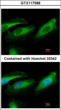
Pirh2 antibodyGTX117588
ApplicationsImmunoFluorescence, Western Blot, ImmunoCytoChemistry
ReactivityHuman
TargetRCHY1
- SizePrice
Product group Antibodies
ApplicationsWestern Blot, ELISA
ReactivityHuman
TargetRCHY1
- SizePrice