

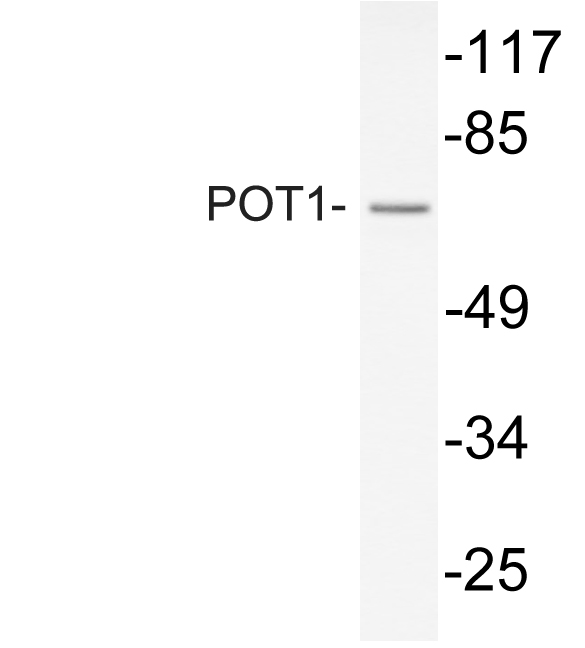
POT1 Rabbit pAb, FITC conjugated
ORB103258
ApplicationsImmunoFluorescence
Product group Antibodies
ReactivityBovine, Equine, Human, Mouse, Porcine, Rat
Overview
- SupplierBiorbyt
- Product NamePOT1 Rabbit pAb, FITC conjugated
- Delivery Days Customer16
- ApplicationsImmunoFluorescence
- Applications SupplierIF=1:100-500 IF
- CertificationResearch Use Only
- ClonalityPolyclonal
- Concentration1 mg/ml
- ConjugateFITC
- HostRabbit
- IsotypeIgG
- Scientific DescriptionPOT1 Rabbit pAb, FITC conjugated
- ReactivityBovine, Equine, Human, Mouse, Porcine, Rat
- Storage Instruction-20°C
- UNSPSC12352203
Related products

Product group Antibodies
POT1 AntibodyCSB-PA003831
ApplicationsWestern Blot, ELISA
ReactivityHuman, Monkey
TargetPOT1
- SizePrice
Product group Antibodies
Anti-POT1 AntibodyA101330
ApplicationsWestern Blot, ELISA
ReactivityHuman
- SizePrice

Product group Antibodies
Anti-POT1 AntibodyHPA068538
ApplicationsImmunoCytoChemistry
ReactivityHuman
TargetPOT1
- SizePrice
Product group Antibodies

POT1 AntibodyLS-C331495
ApplicationsWestern Blot, ImmunoHistoChemistry
ReactivityHuman, Mouse, Rat
TargetPOT1
- SizePrice
Product group Antibodies
ApplicationsImmunoPrecipitation, Western Blot, ImmunoCytoChemistry, ImmunoHistoChemistry
TargetPOT1
- SizePrice

Product group Antibodies
Anti-POT1 Antibody Picoband(r)PB9780-CARRIER-FREE
ApplicationsFlow Cytometry, Western Blot
ReactivityHuman, Mouse, Rat
TargetPOT1
- SizePrice
Product group Antibodies
POT1 antibody [N1C1-2]GTX119700
ApplicationsWestern Blot
ReactivityHuman
TargetPOT1
- SizePrice
Product group Antibodies
Anti-POT1 Antibody144-01491
ApplicationsWestern Blot, ImmunoHistoChemistry
ReactivityHuman, Mouse, Rat
TargetPOT1
- SizePrice