

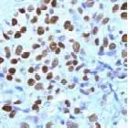
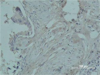
GTX15510 staining Progesterone Receptor in breast carcinoma by Immunohistochemistry (FFPE-sections).
 antibody (GTX15509)")
GTX15510 staining Progesterone Receptor in breast carcinoma by Immunohistochemistry (FFPE-sections).
Progesterone Receptor antibody (ready-to-use)
GTX15510
ApplicationsImmunoHistoChemistry, ImmunoHistoChemistry Paraffin
Product group Antibodies
ReactivityHuman
TargetPGR
Overview
- SupplierGeneTex
- Product NameProgesterone Receptor antibody (ready-to-use)
- Delivery Days Customer9
- Application Supplier NoteIHC-P: Working dilution, ready to use for 10 minutes at RT. Staining of formalin-fixed tissues requires boiling tissue sections in 10mM citrate buffer, pH 6.0 for 10 minutes followed by cooling at RT for 20 minutes. Optimal dilutions/concentrations should be determined by the end user.
- ApplicationsImmunoHistoChemistry, ImmunoHistoChemistry Paraffin
- CertificationResearch Use Only
- ClonalityPolyclonal
- ConjugateUnconjugated
- Gene ID5241
- Target namePGR
- Target descriptionprogesterone receptor
- Target synonymsNR3C3, PR, progesterone receptor, nuclear receptor subfamily 3 group C member 3
- HostRabbit
- IsotypeIgG
- Protein IDP06401
- Protein NameProgesterone receptor
- Scientific DescriptionThis gene encodes a member of the steroid receptor superfamily. The encoded protein mediates the physiological effects of progesterone, which plays a central role in reproductive events associated with the establishment and maintenance of pregnancy. This gene uses two distinct promotors and translation start sites in the first exon to produce several transcript variants, both protein coding and non-protein coding. Two of the isoforms (A and B) are identical except for an additional 165 amino acids found in the N-terminus of isoform B and mediate their own response genes and physiologic effects with little overlap. [provided by RefSeq, Sep 2015]
- ReactivityHuman
- Storage Instruction2°C to 8°C
- UNSPSC41116161
Datasheet
Related products
Product group Antibodies
ApplicationsImmunoFluorescence, ELISA, ImmunoHistoChemistry
ReactivityHuman, Mouse, Rat
- SizePrice
Product group Antibodies

Anti-Progesterone Receptor [14B3]Ab03318-10.0
ApplicationsWestern Blot, ELISA, ImmunoHistoChemistry
ReactivityHuman
TargetPGR
- SizePrice
Product group Antibodies
Anti-PGR Antibody144-61602
ApplicationsWestern Blot, ImmunoHistoChemistry
ReactivityHuman, Mouse, Rat
TargetPGR
- SizePrice

Product group Antibodies
Anti-PGR AntibodyAMAB91529
ApplicationsWestern Blot, ImmunoCytoChemistry, ImmunoHistoChemistry
ReactivityHuman
TargetPGR
- SizePrice
Product group Antibodies
References
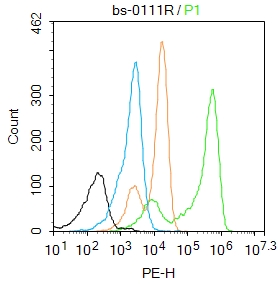
Progesterone Receptor AntibodyBS-0111R
ApplicationsFlow Cytometry, ImmunoFluorescence, Western Blot, ELISA, ImmunoCytoChemistry, ImmunoHistoChemistry, ImmunoHistoChemistry Frozen, ImmunoHistoChemistry Paraffin
ReactivityCanine, Equine, Human, Mouse, Porcine, Rabbit, Rat
TargetPGR
- SizePrice
Product group Antibodies
PGR Monoclonal AntibodyCSB-MA080142
ApplicationsImmunoFluorescence, ELISA, ImmunoHistoChemistry
ReactivityHuman, Mouse, Rat
TargetPGR
- SizePrice
Product group Antibodies
ApplicationsImmunoPrecipitation, Western Blot, ImmunoCytoChemistry, ImmunoHistoChemistry
TargetPGR
- SizePrice
![ICC/IF analysis of untreated T47D cells (left panel) or stimulated cells with 100 nM of Progesterone (right panel) using GTX30164 Progesterone Receptor (phospho Ser190) antibody [1154]. Green : Primary antibody Blue : Nuclei Fixation : Formalin Permeabilization : 0.1% Triton X-100 in TBS for 5-10 minute](https://www.genetex.com/upload/website/prouct_img/normal/GTX30164/GTX30164_723_ICC-IF_w_23060722_451.webp)
Product group Antibodies
ApplicationsImmunoFluorescence, ImmunoPrecipitation, Western Blot, ELISA, ImmunoCytoChemistry, ImmunoHistoChemistry, ImmunoHistoChemistry Paraffin
ReactivityHuman, Mouse
TargetPGR
- SizePrice
![Western blot of whole cell T47D lysate prepared from cells that had been incubated in the presence of the synthetic progestin agonist R5020 (500 nM) showing specific immunolabeling of the ~90k PR-A isoform and the ~120 PR-B isoform of the progesterone receptor phosphorylated at Ser294 using Progesterone Receptor (phospho Ser294) antibody [608] (GTX30165). The immunolabeling is blocked by the phosphopeptide used as the antigen (not shown).](https://www.genetex.com/upload/website/prouct_img/normal/GTX30165/Progesterone-Receptor-phospho-Ser294-antibody-608__GTX30165-1_w_23060722_991.webp)
Product group Antibodies
ApplicationsWestern Blot, ImmunoHistoChemistry
ReactivityHuman
TargetPGR
- SizePrice