

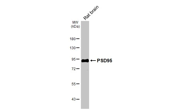
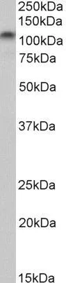
Rat tissue extract (50 μg) was separated by 7.5% SDS-PAGE, and the membrane was blotted with PSD95 antibody [HL3165] (GTX640675) diluted at 1:1000. The HRP-conjugated anti-rabbit IgG antibody (GTX213110-01) was used to detect the primary antibody.
![Various tissue extracts (50 μg) were separated by 7.5% SDS-PAGE, and the membrane was blotted with PSD95 antibody [HL3165] (GTX640675) diluted at 1:100000. The HRP-conjugated anti-rabbit IgG antibody (GTX213110-01) was used to detect the primary antibody.](https://www.genetex.com/upload/website/prouct_img/normal/GTX640675/GTX640675_T-45481_20240809_WB_M_tissue_24081300_465.webp "Various tissue extracts (50 μg) were separated by 7.5% SDS-PAGE, and the membrane was blotted with PSD95 antibody [HL3165] (GTX640675) diluted at 1:100000. The HRP-conjugated anti-rabbit IgG antibody (GTX213110-01) was used to detect the primary antibody.")
![PSD95 antibody [HL3165] detects PSD95 protein by immunohistochemical analysis. Sample: Paraffin-embedded mouse cerebellum. Green: PSD95 stained by PSD95 antibody [HL3165] (GTX640675) diluted at 1:100. Red: beta Tubulin 3/ Tuj1, a neural marker, stained by beta Tubulin 3/ Tuj1 antibody [GT11710] (GTX631836) diluted at 1:500. Blue: Fluoroshield with DAPI (GTX30920). Antigen Retrieval: Citrate buffer, pH 6.0, 15 min](https://www.genetex.com/upload/website/prouct_img/normal/GTX640675/GTX640675_T-45481_20240923_IHC-P_M_24092600_947.webp "PSD95 antibody [HL3165] detects PSD95 protein by immunohistochemical analysis. Sample: Paraffin-embedded mouse cerebellum. Green: PSD95 stained by PSD95 antibody [HL3165] (GTX640675) diluted at 1:100. Red: beta Tubulin 3/ Tuj1, a neural marker, stained by beta Tubulin 3/ Tuj1 antibody [GT11710] (GTX631836) diluted at 1:500. Blue: Fluoroshield with DAPI (GTX30920). Antigen Retrieval: Citrate buffer, pH 6.0, 15 min")
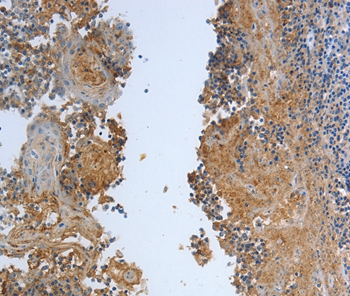
![PSD95 antibody [HL3165] detects PSD95 protein by immunohistochemical analysis. Sample: Paraffin-embedded rat eye. Green: PSD95 stained by PSD95 antibody [HL3165] (GTX640675) diluted at 1:250. Red: beta Tubulin 3/ Tuj1 stained by beta Tubulin 3/ Tuj1 antibody [GT11710] (GTX631836) diluted at 1:500. Blue: Fluoroshield with DAPI (GTX30920). Antigen Retrieval: Citrate buffer, pH 6.0, 15 min](https://www.genetex.com/upload/website/prouct_img/normal/GTX640675/GTX640675_T-45481_20241113_IHC-P_R_24111918_217.webp "PSD95 antibody [HL3165] detects PSD95 protein by immunohistochemical analysis. Sample: Paraffin-embedded rat eye. Green: PSD95 stained by PSD95 antibody [HL3165] (GTX640675) diluted at 1:250. Red: beta Tubulin 3/ Tuj1 stained by beta Tubulin 3/ Tuj1 antibody [GT11710] (GTX631836) diluted at 1:500. Blue: Fluoroshield with DAPI (GTX30920). Antigen Retrieval: Citrate buffer, pH 6.0, 15 min")
![PSD95 antibody [HL3165] detects PSD95 protein by immunohistochemical analysis. Sample: Frozen-sectioned mouse eye. Green: PSD95 stained by PSD95 antibody [HL3165] (GTX640675) diluted at 1:250. Red: beta Tubulin 3/ Tuj1 stained by beta Tubulin 3/ Tuj1 antibody [GT11710] (GTX631836) diluted at 1:500. Blue: Fluoroshield with DAPI (GTX30920).](https://www.genetex.com/upload/website/prouct_img/normal/GTX640675/GTX640675_T-45481_20250117_IHC-Fr_M_25012200_382.webp "PSD95 antibody [HL3165] detects PSD95 protein by immunohistochemical analysis. Sample: Frozen-sectioned mouse eye. Green: PSD95 stained by PSD95 antibody [HL3165] (GTX640675) diluted at 1:250. Red: beta Tubulin 3/ Tuj1 stained by beta Tubulin 3/ Tuj1 antibody [GT11710] (GTX631836) diluted at 1:500. Blue: Fluoroshield with DAPI (GTX30920).")
![Various whole cell extracts (30 μg) were separated by 7.5% SDS-PAGE, and the membrane was blotted with PSD95 antibody [HL3165] (GTX640675) diluted at 1:1000. The HRP-conjugated anti-rabbit IgG antibody (GTX213110-01) was used to detect the primary antibody, and the signal was developed with Trident ECL plus-Enhanced. Corresponding RNA expression data are based on Human Protein Atlas program.](https://www.genetex.com/upload/website/prouct_img/normal/GTX640675/GTX640675_45537_20250418_WB_TPM_watermark_25042420_985.webp "Various whole cell extracts (30 μg) were separated by 7.5% SDS-PAGE, and the membrane was blotted with PSD95 antibody [HL3165] (GTX640675) diluted at 1:1000. The HRP-conjugated anti-rabbit IgG antibody (GTX213110-01) was used to detect the primary antibody, and the signal was developed with Trident ECL plus-Enhanced. Corresponding RNA expression data are based on Human Protein Atlas program.")
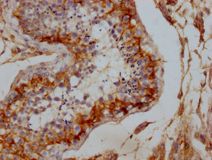
![PSD95 antibody [HL3165] detects PSD95 protein by immunohistochemical analysis. Sample: Paraffin-embedded mouse brain. PSD95 stained by PSD95 antibody [HL3165] (GTX640675) diluted at 1:200. Antigen Retrieval: Citrate buffer, pH 6.0, 15 min](https://www.genetex.com/upload/website/prouct_img/normal/GTX640675/GTX640675_45537_20250926_IHC-P_M_1_25100122_965.webp "PSD95 antibody [HL3165] detects PSD95 protein by immunohistochemical analysis. Sample: Paraffin-embedded mouse brain. PSD95 stained by PSD95 antibody [HL3165] (GTX640675) diluted at 1:200. Antigen Retrieval: Citrate buffer, pH 6.0, 15 min")
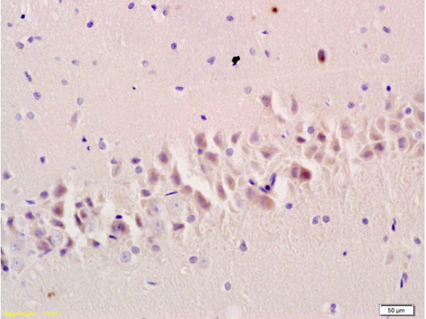
![PSD95 antibody [HL3165] detects PSD95 protein by immunohistochemical analysis. Sample: Paraffin-embedded mouse hippocampus. PSD95 stained by PSD95 antibody [HL3165] (GTX640675) diluted at 1:200. Antigen Retrieval: Citrate buffer, pH 6.0, 15 min](https://www.genetex.com/upload/website/prouct_img/normal/GTX640675/GTX640675_45537_20250926_IHC-P_M_2_25100122_723.webp "PSD95 antibody [HL3165] detects PSD95 protein by immunohistochemical analysis. Sample: Paraffin-embedded mouse hippocampus. PSD95 stained by PSD95 antibody [HL3165] (GTX640675) diluted at 1:200. Antigen Retrieval: Citrate buffer, pH 6.0, 15 min")
Rat tissue extract (50 μg) was separated by 7.5% SDS-PAGE, and the membrane was blotted with PSD95 antibody [HL3165] (GTX640675) diluted at 1:1000. The HRP-conjugated anti-rabbit IgG antibody (GTX213110-01) was used to detect the primary antibody.
PSD95 antibody [HL3165]
GTX640675
ApplicationsWestern Blot, ImmunoHistoChemistry, ImmunoHistoChemistry Frozen, ImmunoHistoChemistry Paraffin
Product group Antibodies
ReactivityHuman, Mouse, Rat
TargetDLG4
Overview
- SupplierGeneTex
- Product NamePSD95 antibody [HL3165]
- Delivery Days Customer7
- Application Supplier NoteWB: 1:500-1:3000. *Optimal dilutions/concentrations should be determined by the researcher.Not tested in other applications.
- ApplicationsWestern Blot, ImmunoHistoChemistry, ImmunoHistoChemistry Frozen, ImmunoHistoChemistry Paraffin
- CertificationResearch Use Only
- ClonalityMonoclonal
- Clone IDHL3165
- Concentration1 mg/ml
- ConjugateUnconjugated
- Gene ID1742
- Target nameDLG4
- Target descriptiondiscs large MAGUK scaffold protein 4
- Target synonymsMRD62, PSD95, SAP-90, SAP90, disks large homolog 4, Tax interaction protein 15, discs large homolog 4, post-synaptic density protein 95, synapse-associated protein 90
- HostRabbit
- IsotypeIgG
- Protein IDP78352
- Protein NameDisks large homolog 4
- Scientific DescriptionThis gene encodes a member of the membrane-associated guanylate kinase (MAGUK) family. It heteromultimerizes with another MAGUK protein, DLG2, and is recruited into NMDA receptor and potassium channel clusters. These two MAGUK proteins may interact at postsynaptic sites to form a multimeric scaffold for the clustering of receptors, ion channels, and associated signaling proteins. Multiple transcript variants encoding different isoforms have been found for this gene. [provided by RefSeq, Jul 2008]
- ReactivityHuman, Mouse, Rat
- Storage Instruction-20°C or -80°C,2°C to 8°C
- UNSPSC41116161
Datasheet
Related products
Product group Antibodies
Anti-DLG4 AntibodyA46884
ApplicationsImmunoHistoChemistry
ReactivityHuman, Mouse, Rat
- SizePrice
Product group Antibodies

Anti-PSD-95 [K28/43]Ab02108-10.0
ApplicationsImmunoFluorescence, ImmunoPrecipitation, Western Blot
ReactivityHuman, Mouse, Rat
TargetDLG4
- SizePrice
Product group Antibodies
Anti-DLG4 Antibody144-07889
ApplicationsWestern Blot, ImmunoHistoChemistry
ReactivityHuman, Mouse, Rat
TargetDLG4
- SizePrice

Product group Antibodies
Anti-PSD95/DLG4 Antibody Picoband(r)A02128-CARRIER-FREE
ApplicationsFlow Cytometry, Western Blot, ELISA
ReactivityHuman, Mouse, Rat
TargetDLG4
- SizePrice
Product group Antibodies
References
PSD95 Polyclonal AntibodyBS-0179R
ApplicationsFlow Cytometry, ImmunoFluorescence, Western Blot, ELISA, ImmunoCytoChemistry, ImmunoHistoChemistry, ImmunoHistoChemistry Frozen, ImmunoHistoChemistry Paraffin
ReactivityBovine, Canine, Equine, Human, Mouse, Porcine, Rabbit, Rat, Sheep
TargetDLG4
- SizePrice
Product group Antibodies
DLG4 AntibodyCSB-PA006938LA01HU
ApplicationsELISA, ImmunoHistoChemistry
ReactivityHuman
TargetDLG4
- SizePrice
Product group Antibodies

Goat anti-DLG4 / PSD95EB09300
ApplicationsWestern Blot, ELISA
ReactivityBovine, Canine, Human, Mouse, Rat
TargetDLG4
- SizePrice
Product group Antibodies
Dlg4 Recombinant AntibodyCAC12531
ApplicationsWestern Blot, ELISA
ReactivityMouse, Rat
TargetDLG4
- SizePrice
Product group Antibodies

DLG4 / PSD95 AntibodyLS-C406001
ApplicationsELISA, ImmunoHistoChemistry
ReactivityHuman, Mouse, Rat
TargetDLG4
- SizePrice
Product group Antibodies
References
ApplicationsWestern Blot
ReactivityHuman, Rat
TargetDLG4
- SizePrice