

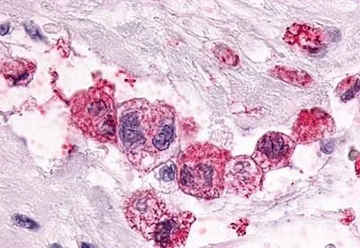
IHC-P analysis of human pancreas, carcinoma tissue using GTX13381 PTGER3 antibody. Antigen retrieval : Heat-induced antigen retrieval


IHC-P analysis of human pancreas, carcinoma tissue using GTX13381 PTGER3 antibody. Antigen retrieval : Heat-induced antigen retrieval
PTGER3 antibody
GTX13381
ApplicationsImmunoHistoChemistry, ImmunoHistoChemistry Paraffin
Product group Antibodies
ReactivityHuman, Mammals, Monkey
TargetPTGER3
Overview
- SupplierGeneTex
- Product NamePTGER3 antibody
- Delivery Days Customer9
- Application Supplier NoteIHC-P: 1 - 3 microg/ml. IHC-P: 1 - 3 microg/ml. *Optimal dilutions/concentrations should be determined by the researcher.Not tested in other applications.
- ApplicationsImmunoHistoChemistry, ImmunoHistoChemistry Paraffin
- CertificationResearch Use Only
- ClonalityPolyclonal
- Concentration1 mg/ml
- ConjugateUnconjugated
- Gene ID5733
- Target namePTGER3
- Target descriptionprostaglandin E receptor 3
- Target synonymsEP3, EP3-I, EP3-II, EP3-III, EP3-IV, EP3-VI, EP3e, PGE2-R, lnc003875, prostaglandin E2 receptor EP3 subtype, PGE receptor, EP3 subtype, PGE2 receptor EP3 subtype, prostaglandin E receptor 3 (subtype EP3), prostaglandin receptor (PGE-2), prostanoid EP3 receptor
- HostRabbit
- IsotypeIgG
- Protein IDP43115
- Protein NameProstaglandin E2 receptor EP3 subtype
- Scientific DescriptionThe protein encoded by this gene is a member of the G-protein coupled receptor family. This protein is one of four receptors identified for prostaglandin E2 (PGE2). This receptor may have many biological functions, which involve digestion, nervous system, kidney reabsorption, and uterine contraction activities. Studies of the mouse counterpart suggest that this receptor may also mediate adrenocorticotropic hormone response as well as fever generation in response to exogenous and endogenous stimuli. Multiple transcript variants encoding different isoforms have been found for this gene. [provided by RefSeq, Aug 2009]
- ReactivityHuman, Mammals, Monkey
- Storage Instruction-20°C or -80°C,2°C to 8°C
- UNSPSC41116161
Datasheet
Related products

Product group Antibodies
Anti-PTGER3 AntibodyA100334
ApplicationsImmunoFluorescence, ELISA
ReactivityHuman
- SizePrice

Product group Antibodies
Anti-PTGER3 Antibody Picoband(r)A02548-1-CARRIER-FREE
ApplicationsFlow Cytometry, Western Blot, ELISA
ReactivityHuman
TargetPTGER3
- SizePrice
Product group Antibodies
Anti-PTGER3 Antibody144-65763
ApplicationsWestern Blot
ReactivityHuman, Mouse, Rat
TargetPTGER3
- SizePrice
Product group Antibodies
EP3/PTGER3 Polyclonal AntibodyBS-1876R
ApplicationsFlow Cytometry, Western Blot, ELISA, ImmunoHistoChemistry, ImmunoHistoChemistry Paraffin
ReactivityMouse, Rat
TargetPTGER3
- SizePrice
Product group Antibodies
PTGER3 AntibodyCSB-PA002352
ApplicationsImmunoFluorescence, Western Blot, ELISA
ReactivityHuman
TargetPTGER3
- SizePrice
Product group Antibodies

Goat anti-PTGER3 / EP3EB08205
ApplicationsWestern Blot, ELISA
ReactivityHuman
TargetPTGER3
- SizePrice
Product group Antibodies
ApplicationsWestern Blot, ELISA, ImmunoCytoChemistry, ImmunoHistoChemistry, ImmunoHistoChemistry Frozen, ImmunoHistoChemistry Paraffin
ReactivityMouse
TargetPTGER3
- SizePrice
Product group Antibodies

PTGER3 / EP3 AntibodyLS-C408750
ApplicationsWestern Blot, ImmunoHistoChemistry
ReactivityHuman, Mouse, Rat
TargetPTGER3
- SizePrice
![Prostaglandin E Receptor EP3 antibody [5F5]](https://www.genetex.com/upload/website/prouct_img/normal/GTX16152/img25886_w_23060620_363.webp)
Product group Antibodies
PTGER3 antibody [5F5]GTX16152
ApplicationsWestern Blot
ReactivityBovine, Human, Rat
TargetPTGER3
- SizePrice
Product group Antibodies
Anti-PTGER3 AntibodyHPA010689
ApplicationsImmunoHistoChemistry
ReactivityHuman
TargetPTGER3
- SizePrice