

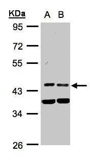
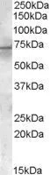
Sample(30 ug whole cell lysate) A:HeLa S3(GTX14654) B:MOLT4 (GTX27912) 10% SDS PAGE GTX105999 diluted at 1:1000
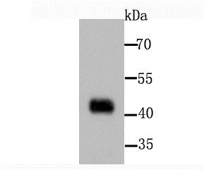
![PDK1 antibody [N1C3] detects PDK1 protein by western blot analysis. A. 30 μg 293T whole cell extract B. 30 μg whole cell extract of human PDK1 transfected 293T cells 10 % SDS-PAGE PDK1 antibody [N1C3] (GTX105999) dilution: 1:10000](https://www.genetex.com/upload/website/prouct_img/normal/GTX105999/GTX105999_39953_WB_B_w_23060120_911.webp "PDK1 antibody [N1C3] detects PDK1 protein by western blot analysis. A. 30 μg 293T whole cell extract B. 30 μg whole cell extract of human PDK1 transfected 293T cells 10 % SDS-PAGE PDK1 antibody [N1C3] (GTX105999) dilution: 1:10000")
Sample(30 ug whole cell lysate) A:HeLa S3(GTX14654) B:MOLT4 (GTX27912) 10% SDS PAGE GTX105999 diluted at 1:1000
Pyruvate dehydrogenase kinase 1 / PDK1 antibody [N1C3]
GTX105999
ApplicationsWestern Blot
Product group Antibodies
ReactivityHuman
TargetPDK1
Overview
- SupplierGeneTex
- Product NamePyruvate dehydrogenase kinase 1 / PDK1 antibody [N1C3]
- Delivery Days Customer9
- Application Supplier NoteWB: 1:500-1:20000. *Optimal dilutions/concentrations should be determined by the researcher.Not tested in other applications.
- ApplicationsWestern Blot
- CertificationResearch Use Only
- ClonalityPolyclonal
- Concentration1 mg/ml
- ConjugateUnconjugated
- Gene ID5163
- Target namePDK1
- Target descriptionpyruvate dehydrogenase kinase 1
- Target synonyms[Pyruvate dehydrogenase (acetyl-transferring)] kinase isozyme 1, mitochondrial, pyruvate dehydrogenase (acetyl-transferring) kinase isozyme 1, mitochondrial, PDH kinase 1, mitochondrial pyruvate dehydrogenase, lipoamide, kinase isoenzyme 1, pyruvate dehydrogenase kinase, isoenzyme 1
- HostRabbit
- IsotypeIgG
- Protein IDQ15118
- Protein Name[Pyruvate dehydrogenase (acetyl-transferring)] kinase isozyme 1, mitochondrial
- Scientific DescriptionPyruvate dehydrogenase (PDH) is a mitochondrial multienzyme complex that catalyzes the oxidative decarboxylation of pyruvate and is one of the major enzymes responsible for the regulation of homeostasis of carbohydrate fuels in mammals. The enzymatic activity is regulated by a phosphorylation/dephosphorylation cycle. Phosphorylation of PDH by a specific pyruvate dehydrogenase kinase (PDK) results in inactivation. [provided by RefSeq]
- ReactivityHuman
- Storage Instruction-20°C or -80°C,2°C to 8°C
- UNSPSC41116161
Datasheet
Related products
Product group Antibodies
Anti-PDK1 AntibodyA96144
ApplicationsWestern Blot, ELISA, ImmunoHistoChemistry
ReactivityHuman, Mouse, Rat
- SizePrice

Product group Antibodies
Anti-Mitochondrial Pyruvate dehydrogenase kinase 1/PDK1 Antibody Picoband(r)A01268-1-CARRIER-FREE
ApplicationsWestern Blot, ELISA, ImmunoHistoChemistry
ReactivityHuman, Rat
TargetPDK1
- SizePrice
Product group Antibodies
Anti-PDK1 Antibody144-61653
ApplicationsWestern Blot
ReactivityHuman, Mouse, Rat
TargetPDK1
- SizePrice
Product group Antibodies
PDPK1 Recombinant AntibodyBSM-54037R
ApplicationsFlow Cytometry, ImmunoFluorescence, Western Blot, ImmunoCytoChemistry, ImmunoHistoChemistry, ImmunoHistoChemistry Frozen, ImmunoHistoChemistry Paraffin
ReactivityHuman, Mouse, Rat
TargetPDK1
- SizePrice
Product group Antibodies
PDK1 AntibodyCSB-PA003729
ApplicationsWestern Blot, ELISA
ReactivityHuman, Mouse, Rat
TargetPDK1
- SizePrice
Product group Antibodies

Goat anti-PDK1EB06983
ApplicationsWestern Blot, ELISA
ReactivityBovine, Canine, Human, Mouse, Porcine, Rat
TargetPDK1
- SizePrice
Product group Antibodies
Pdk1 Polyclonal AntibodyCAC11151
ApplicationsWestern Blot, ELISA, ImmunoHistoChemistry
ReactivityMouse
TargetPDK1
- SizePrice
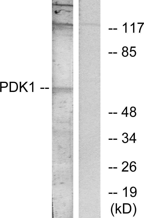
![Western blot using GeneTex's affinity purified anti-PDK-1 antibody shows detection of myc-tagged human PDK-1 at 60kDa in ~10 μg of a virus infected Sf9 cell lysate (arrow). The nitrocellulose membrane was probed overnight at 4o C with the primary antibody diluted 1:750 in 1% non-fat dry milk. HRP conjugated Goat-anti-Rabbit IgG [H&L] and chemiluminescent detection is recommended. Other detection systems will yield similar results.](https://www.genetex.com/upload/website/prouct_img/normal/GTX22495/GTX22495_20160330_WB_w_23060620_134.webp)
Product group Antibodies
ApplicationsWestern Blot, ELISA, ImmunoHistoChemistry
ReactivityHuman
TargetPDK1
- SizePrice
Product group Antibodies

PDK1 AntibodyLS-C331125
ApplicationsWestern Blot
ReactivityHuman, Mouse, Rat
TargetPDK1
- SizePrice
Product group Antibodies
ApplicationsWestern Blot
ReactivityHuman, Rat
TargetPDK1
- SizePrice