

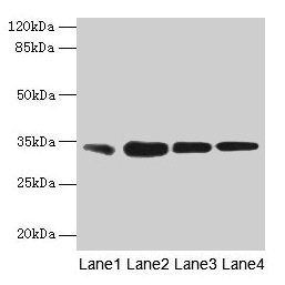
Western blot All lanes: RAD23A antibody at 3microg/ml Lane 1: MCF-7 whole cell lysate Lane 2: Hela whole cell lysate Lane 3: Raji whole cell lysate Lane 4: Jurkat whole cell lysate Secondary Goat polyclonal to rabbit IgG at 1/10000 dilution Predicted band size: 40, 34 kDa Observed band size: 34 kDa

Western blot All lanes: RAD23A antibody at 3microg/ml Lane 1: MCF-7 whole cell lysate Lane 2: Hela whole cell lysate Lane 3: Raji whole cell lysate Lane 4: Jurkat whole cell lysate Secondary Goat polyclonal to rabbit IgG at 1/10000 dilution Predicted band size: 40, 34 kDa Observed band size: 34 kDa
RAD23A Antibody
CSB-PA03774A0RB
ApplicationsWestern Blot, ELISA, ImmunoHistoChemistry
Product group Antibodies
ReactivityHuman
TargetRAD23A
Overview
- SupplierCusabio
- Product NameRAD23A Antibody
- Delivery Days Customer20
- ApplicationsWestern Blot, ELISA, ImmunoHistoChemistry
- CertificationResearch Use Only
- ClonalityPolyclonal
- ConjugateUnconjugated
- Gene ID5886
- Target nameRAD23A
- Target descriptionRAD23 nucleotide excision repair protein A
- Target synonymsHHR23A, HR23A, UV excision repair protein RAD23 homolog A, RAD23 homolog A, nucleotide excision repair protein, RAD23, yeast homolog, A
- HostRabbit
- IsotypeIgG
- Protein IDP54725
- Protein NameUV excision repair protein RAD23 homolog A
- Scientific DescriptionMultiubiquitin chain receptor involved in modulation of proteasomal degradation. Binds to Lys-48-linked polyubiquitin chains in a length-dependent manner and with a lower affinity to Lys-63-linked polyubiquitin chains. Proposed to be capable to bind simultaneously to the 26S proteasome and to polyubiquitinated substrates and to deliver ubiquitinated proteins to the proteasome. Involved in nucleotide excision repair and is thought to be functional equivalent for RAD23B in global genome nucleotide excision repair (GG-NER) by association with XPC. In vitro, the XPC:RAD23A dimer has NER activity. Can stabilize XPC. (Microbial infection) Involved in Vpr-dependent replication of HIV-1 in non-proliferating cells and primary macrophages. Required for the association of HIV-1 Vpr with the host proteasome.
- ReactivityHuman
- Storage Instruction-20°C or -80°C
- UNSPSC41116161
Related products

Product group Antibodies
Anti-RAD23A AntibodyA98763
ApplicationsWestern Blot, ELISA
ReactivityHuman, Mouse
- SizePrice

Product group Antibodies
Anti-hHR23A/RAD23A Antibody Picoband(r)A03243-3-CARRIER-FREE
ApplicationsFlow Cytometry, Western Blot
ReactivityHuman, Mouse, Rat
TargetRAD23A
- SizePrice
Product group Antibodies
RAD23A Recombinant AntibodyBSM-62209R
ApplicationsFlow Cytometry, Western Blot
ReactivityHuman, Mouse, Rat
TargetRAD23A
- SizePrice
Product group Antibodies
RAD23A Polyclonal AntibodyCAC14049
ApplicationsWestern Blot, ELISA, ImmunoHistoChemistry
TargetRAD23A
- SizePrice
Product group Antibodies
RAD23A antibody [N1N2], N-termGTX100425
ApplicationsImmunoFluorescence, ImmunoCytoChemistry, ImmunoHistoChemistry, ImmunoHistoChemistry Frozen
ReactivityHuman, Mouse
TargetRAD23A
- SizePrice
Product group Antibodies

RAD23A / HHR23A AntibodyLS-C332409
ApplicationsWestern Blot, ImmunoHistoChemistry
ReactivityHuman, Mouse
TargetRAD23A
- SizePrice

Product group Antibodies
Anti-RAD23A AntibodyHPA026418
ApplicationsWestern Blot, ImmunoCytoChemistry
ReactivityHuman
TargetRAD23A
- SizePrice
Product group Antibodies
ApplicationsImmunoFluorescence, Western Blot, ELISA, ImmunoCytoChemistry
ReactivityHuman
TargetRAD23A
- SizePrice