

RAD23A antibody [N1N2], N-term
GTX100425
ApplicationsImmunoFluorescence, ImmunoCytoChemistry, ImmunoHistoChemistry, ImmunoHistoChemistry Frozen
Product group Antibodies
ReactivityHuman, Mouse
TargetRAD23A
Overview
- SupplierGeneTex
- Product NameRAD23A antibody [N1N2], N-term
- Delivery Days Customer9
- ApplicationsImmunoFluorescence, ImmunoCytoChemistry, ImmunoHistoChemistry, ImmunoHistoChemistry Frozen
- CertificationResearch Use Only
- ClonalityPolyclonal
- Concentration6.1 mg/ml
- ConjugateUnconjugated
- Gene ID5886
- Target nameRAD23A
- Target descriptionRAD23 nucleotide excision repair protein A
- Target synonymsHHR23A, HR23A, UV excision repair protein RAD23 homolog A, RAD23 homolog A, nucleotide excision repair protein, RAD23, yeast homolog, A
- HostRabbit
- IsotypeIgG
- Protein IDP54725
- Protein NameUV excision repair protein RAD23 homolog A
- Scientific DescriptionThe protein encoded by this gene is one of two human homologs of Saccharomyces cerevisiae Rad23, a protein involved in nucleotide excision repair (NER). This protein was shown to interact with, and elevate the nucleotide excision activity of 3-methyladenine-DNA glycosylase (MPG), which suggested a role in DNA damage recognition in base excision repair. This protein contains an N-terminal ubiquitin-like domain, which was reported to interact with 26S proteasome, as well as with ubiquitin protein ligase E6AP, and thus suggests that this protein may be involved in the ubiquitin mediated proteolytic pathway in cells. [provided by RefSeq]
- ReactivityHuman, Mouse
- Storage Instruction-20°C or -80°C,2°C to 8°C
- UNSPSC41116161
Datasheet
Related products

Product group Antibodies
Anti-RAD23A AntibodyA98763
ApplicationsWestern Blot, ELISA
ReactivityHuman, Mouse
- SizePrice

Product group Antibodies
Anti-hHR23A/RAD23A Antibody Picoband(r)A03243-3-CARRIER-FREE
ApplicationsFlow Cytometry, Western Blot
ReactivityHuman, Mouse, Rat
TargetRAD23A
- SizePrice
Product group Antibodies
RAD23A Recombinant AntibodyBSM-62209R
ApplicationsFlow Cytometry, Western Blot
ReactivityHuman, Mouse, Rat
TargetRAD23A
- SizePrice
Product group Antibodies
RAD23A Polyclonal AntibodyCAC14049
ApplicationsWestern Blot, ELISA, ImmunoHistoChemistry
TargetRAD23A
- SizePrice
Product group Antibodies
RAD23A AntibodyCSB-PA03774A0RB
ApplicationsWestern Blot, ELISA, ImmunoHistoChemistry
ReactivityHuman
TargetRAD23A
- SizePrice
Product group Antibodies
RAD23A antibodyGTX23836
ApplicationsWestern Blot, ELISA
ReactivityHuman
TargetRAD23A
- SizePrice
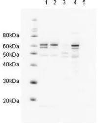
![Various whole cell extracts (30 μg) were separated by 10% SDS-PAGE, and the membrane was blotted with Rad23A antibody [N3C3] (GTX102032) diluted at 1:1000.](https://www.genetex.com/upload/website/prouct_img/normal/GTX102032/GTX102032_39771_20161013_WB_M_w_23060100_350.webp)
Product group Antibodies
RAD23A antibody [N3C3]GTX102032
ApplicationsWestern Blot
ReactivityHuman, Mouse, Rat
TargetRAD23A
- SizePrice
Product group Antibodies

RAD23A / HHR23A AntibodyLS-C332409
ApplicationsWestern Blot, ImmunoHistoChemistry
ReactivityHuman, Mouse
TargetRAD23A
- SizePrice

Product group Antibodies
Anti-RAD23A AntibodyHPA026418
ApplicationsWestern Blot, ImmunoCytoChemistry
ReactivityHuman
TargetRAD23A
- SizePrice
Product group Antibodies
ApplicationsImmunoFluorescence, Western Blot, ELISA, ImmunoCytoChemistry
ReactivityHuman
TargetRAD23A
- SizePrice