

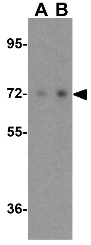
WB analysis of A431 cell lysate using GTX85101 Raf1 antibody. Working concentration : (A) 0.5 and (B) 1 μg/ml
WB analysis of A431 cell lysate using GTX85101 Raf1 antibody. Working concentration : (A) 0.5 and (B) 1 μg/ml
Raf1 antibody
GTX85101
ApplicationsWestern Blot, ELISA
Product group Antibodies
ReactivityHuman, Mouse, Rat
TargetRAF1
Overview
- SupplierGeneTex
- Product NameRaf1 antibody
- Delivery Days Customer9
- Application Supplier NoteWB: 0.5 - 1 microg/mL. *Optimal dilutions/concentrations should be determined by the researcher.Not tested in other applications.
- ApplicationsWestern Blot, ELISA
- CertificationResearch Use Only
- ClonalityPolyclonal
- Concentration1 mg/ml
- ConjugateUnconjugated
- Gene ID5894
- Target nameRAF1
- Target descriptionRaf-1 proto-oncogene, serine/threonine kinase
- Target synonymsCMD1NN, CRAF, NS5, Raf-1, c-Raf, RAF proto-oncogene serine/threonine-protein kinase, C-Raf proto-oncogene, serine/threonine kinase, Oncogene RAF1, proto-oncogene c-RAF, raf proto-oncogene serine/threonine protein kinase, v-raf-1 murine leukemia viral oncogene homolog 1, v-raf-1 murine leukemia viral oncogene-like protein 1
- HostChicken
- IsotypeIgY
- Protein IDP04049
- Protein NameRAF proto-oncogene serine/threonine-protein kinase
- Scientific DescriptionC-raf is the cellular homolog of viral raf gene (v-raf) and encodes a MAP kinase kinase kinase (MAP3K), which functions downstream of the Ras family of membrane associated GTPases. Once activated, C-raf phosphorylates and activates the protein kinases MEK1 and MEK2, which in turn phosphorylate and activate the serine/threonine specific protein kinases, ERK1 and ERK2. These activated ERKs are pleiotropic effectors of cell physiology and play an important role in the control of gene expression involved in the cell division cycle, apoptosis, cell differentiation and cell migration. Mutations in this gene are associated with Noonan syndrome 5 and LEOPARD syndrome 2.
- ReactivityHuman, Mouse, Rat
- Storage Instruction-20°C or -80°C,2°C to 8°C
- UNSPSC12352203
Datasheet
Related products
Product group Antibodies

C-RAF (Phospho-Tyr341) AntibodyABX012449
ApplicationsWestern Blot, ELISA, ImmunoHistoChemistry
- SizePrice
Product group Antibodies
Anti-RAF1 Antibody144-00223
ApplicationsImmunoFluorescence, Western Blot, ImmunoHistoChemistry
ReactivityHuman, Mouse, Rat
TargetRAF1
- SizePrice

Product group Antibodies
Anti-Raf1 Antibody Picoband(r)A00446-1-CARRIER-FREE
ApplicationsFlow Cytometry, Western Blot
ReactivityHuman, Mouse, Rat
TargetRAF1
- SizePrice
![WB analysis of C6 cells were treated by Forskolin (10 uM) at 37oC for 1 hour after serum-starvation overnight using GTX03221 Raf1 (phospho Ser259) antibody [GT1309]. Dilution : 1:1000 Loading : 25μg per lane](https://www.genetex.com/upload/website/prouct_img/normal/GTX03221/GTX03221_81_WB_w_23053123_624.webp)
Product group Antibodies
ApplicationsWestern Blot
ReactivityHuman, Rat
TargetRAF1
- SizePrice
![Raf1 antibody [N3C3] detects Raf1 protein at cell membrane and cytoplasm by immunohistochemical analysis. Sample: Paraffin-embedded human endometrial carcinoma. Raf1 stained by Raf1 antibody [N3C3] (GTX107763) diluted at 1:500. Antigen Retrieval: Citrate buffer, pH 6.0, 15 min](https://www.genetex.com/upload/website/prouct_img/normal/GTX107763/GTX107763_43579_20190628_IHC-P_w_23060120_387.webp)
Product group Antibodies
References
Raf1 antibody [N3C3]GTX107763
ApplicationsImmunoFluorescence, Western Blot, ImmunoCytoChemistry, ImmunoHistoChemistry, ImmunoHistoChemistry Paraffin
ReactivityHuman, Mouse
TargetRAF1
- SizePrice
![Various whole cell extracts (30 μg) were separated by 7.5% SDS-PAGE, and the membrane was blotted with Raf1 antibody [N1C1-2] (GTX111588) diluted at 1:1000. The HRP-conjugated anti-rabbit IgG antibody (GTX213110-01) was used to detect the primary antibody.](https://www.genetex.com/upload/website/prouct_img/normal/GTX111588/GTX111588_40051_20170825_WB_w_23060500_444.webp)
Product group Antibodies
References
Raf1 antibody [N1C1-2]GTX111588
ApplicationsImmunoFluorescence, Western Blot, ImmunoCytoChemistry, ImmunoHistoChemistry, ImmunoHistoChemistry Paraffin
ReactivityHuman, Mouse
TargetRAF1
- SizePrice
![FACS analysis of HeLa cells using GTX60556 Raf1 antibody [4G4]. Green : Raf1 Red : negative control](https://www.genetex.com/upload/website/prouct_img/normal/GTX60556/GTX60556_20170912_FACS_w_23061123_857.webp)
Product group Antibodies
Raf1 antibody [4G4]GTX60556
ApplicationsFlow Cytometry, Western Blot, ELISA, ImmunoHistoChemistry, ImmunoHistoChemistry Paraffin
ReactivityHuman, Monkey, Rat
TargetRAF1
- SizePrice
![WB analysis of HeLa (1), A431 (2), HepG (3), and SW620 (4)cell lysate using GTX60561 Raf1 antibody [4G4].](https://www.genetex.com/upload/website/prouct_img/normal/GTX60561/GTX60561_20170912_WB_w_23061123_821.webp)
Product group Antibodies
Raf1 antibody [4G4]GTX60561
ApplicationsFlow Cytometry, Western Blot, ELISA, ImmunoHistoChemistry, ImmunoHistoChemistry Paraffin
ReactivityHuman
TargetRAF1
- SizePrice
Product group Antibodies
Raf1 (phospho Ser259) antibodyGTX78955
ApplicationsWestern Blot, ImmunoHistoChemistry, ImmunoHistoChemistry Paraffin
ReactivityHuman
TargetRAF1
- SizePrice