

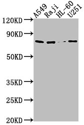
Western Blot Positive WB detected in: A549 whole cell lysate, Raji whole cell lysate, HL60 whole cell lysate, U251 whole cell lysate All lanes: RANBP9 antibody at 1:1000 Secondary Goat polyclonal to rabbit IgG at 1/50000 dilution Predicted band size: 78, 44, 56 kDa Observed band size: 78 kDa
. Section was blocked with 10% normal goat serum 30min at RT. Then primary antibody (1% BSA) was incubated at 4°C overnight. The primary is detected by a biotinylated secondary antibody and visualized using an HRP conjugated SP system.")
Western Blot Positive WB detected in: A549 whole cell lysate, Raji whole cell lysate, HL60 whole cell lysate, U251 whole cell lysate All lanes: RANBP9 antibody at 1:1000 Secondary Goat polyclonal to rabbit IgG at 1/50000 dilution Predicted band size: 78, 44, 56 kDa Observed band size: 78 kDa
RANBP9 Antibody
CSB-PA853501OA01HU
ApplicationsWestern Blot, ELISA, ImmunoHistoChemistry
Product group Antibodies
ReactivityHuman
TargetRANBP9
Overview
- SupplierCusabio
- Product NameRANBP9 Antibody
- Delivery Days Customer20
- ApplicationsWestern Blot, ELISA, ImmunoHistoChemistry
- CertificationResearch Use Only
- ClonalityPolyclonal
- ConjugateUnconjugated
- Gene ID10048
- Target nameRANBP9
- Target descriptionRAN binding protein 9
- Target synonymsBPM-L, BPM90, RANBPM, RanBP7, ran-binding protein 9, Ran Binding Protein in the Microtubule organizing center, novel centrosomal protein RanBPM, ran binding protein, centrosomal, ran-binding protein M
- HostRabbit
- IsotypeIgG
- Protein IDQ96S59
- Protein NameRan-binding protein 9
- Scientific DescriptionMay act as an adapter protein to couple membrane receptors to intracellular signaling pathways. May be involved in signaling of ITGB2/LFA-1 and other integrins. Enhances HGF-MET signaling by recruiting Sos and activating the Ras pathway. Enhances dihydrotestosterone-induced transactivation activity of AR, as well as dexamethasone-induced transactivation activity of NR3C1, but not affect estrogen-induced transactivation. Stabilizes TP73 isoform Alpha, probably by inhibiting its ubiquitination, and increases its proapoptotic activity. Inhibits the kinase activity of DYRK1A and DYRK1B. Inhibits FMR1 binding to RNA (By similarity).
- ReactivityHuman
- Storage Instruction-20°C or -80°C
- UNSPSC41116161
Related products

Product group Antibodies
Anti-RANBP9 Antibody Picoband(r)A03448-1-CARRIER-FREE
ApplicationsFlow Cytometry, ImmunoFluorescence, Western Blot, ELISA, ImmunoCytoChemistry
ReactivityHuman
TargetRANBP9
- SizePrice
Product group Antibodies

RANBPM AntibodyABX430686
ApplicationsImmunoFluorescence, ELISA, ImmunoCytoChemistry, ImmunoHistoChemistry
- SizePrice
Product group Antibodies
Anti-RanBP9 AntibodyA32531
ApplicationsImmunoPrecipitation, Western Blot, ImmunoCytoChemistry
ReactivityHuman
- SizePrice
Product group Antibodies

RANBPM AntibodyLS-C747286
ApplicationsWestern Blot
ReactivityHuman
TargetRANBP9
- SizePrice
Product group Antibodies

Goat anti-RANBPM / RANBP9EB06028
ApplicationsELISA, ImmunoCytoChemistry, ImmunoHistoChemistry
ReactivityCanine, Human, Mouse, Porcine
TargetRANBP9
- SizePrice

Product group Antibodies
Anti-RANBP9 AntibodyHPA050007
ApplicationsImmunoHistoChemistry
ReactivityHuman
TargetRANBP9
- SizePrice
Product group Antibodies
RANBP9 Polyclonal AntibodyCAC15798
ApplicationsWestern Blot, ELISA, ImmunoHistoChemistry
TargetRANBP9
- SizePrice
Product group Antibodies
RanBP9 antibodyGTX106378
ApplicationsWestern Blot
ReactivityHuman, Mouse
TargetRANBP9
- SizePrice
Product group Antibodies
RANBP9 Recombinant Antibody, AbBy Fluor-405 ConjugatedBSM-62362R-BF405
ApplicationsWestern Blot
ReactivityHuman, Mouse, Rat
TargetRANBP9
- SizePrice
Product group Antibodies
Anti-RANBP9 Antibody144-12097
ApplicationsWestern Blot
ReactivityHuman
TargetRANBP9
- SizePrice