

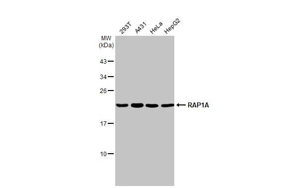
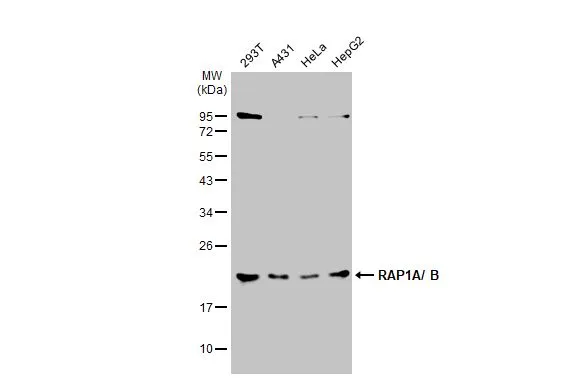
Various whole cell extracts (30 μg) were separated by 12% SDS-PAGE, and the membrane was blotted with RAP1A antibody [HL2415] (GTX638637) diluted at 1:1000. The HRP-conjugated anti-rabbit IgG antibody (GTX213110-01) was used to detect the primary antibody.
Various whole cell extracts (30 μg) were separated by 12% SDS-PAGE, and the membrane was blotted with RAP1A antibody [HL2415] (GTX638637) diluted at 1:1000. The HRP-conjugated anti-rabbit IgG antibody (GTX213110-01) was used to detect the primary antibody.
RAP1A antibody [HL2415]
GTX638637
ApplicationsWestern Blot
Product group Antibodies
ReactivityHuman
TargetRAP1A
Overview
- SupplierGeneTex
- Product NameRAP1A antibody [HL2415]
- Delivery Days Customer9
- Application Supplier NoteWB: 1:500-1:3000. *Optimal dilutions/concentrations should be determined by the researcher.Not tested in other applications.
- ApplicationsWestern Blot
- CertificationResearch Use Only
- ClonalityMonoclonal
- Clone IDHL2415
- Concentration1 mg/ml
- ConjugateUnconjugated
- Gene ID5906
- Target nameRAP1A
- Target descriptionRAP1A, member of RAS oncogene family
- Target synonymsC21KG, G-22K, KREV-1, KREV1, RAP1, SMGP21, ras-related protein Rap-1A, GTP-binding protein smg p21A, Ras-related protein Krev-1
- HostRabbit
- IsotypeIgG
- Protein IDP62834
- Protein NameRas-related protein Rap-1A
- Scientific DescriptionThis gene encodes a member of the Ras family of small GTPases. The encoded protein undergoes a change in conformational state and activity, depending on whether it is bound to GTP or GDP. This protein is activated by several types of guanine nucleotide exchange factors (GEFs), and inactivated by two groups of GTPase-activating proteins (GAPs). The activation status of the encoded protein is therefore affected by the balance of intracellular levels of GEFs and GAPs. The encoded protein regulates signaling pathways that affect cell proliferation and adhesion, and may play a role in tumor malignancy. Pseudogenes of this gene have been defined on chromosomes 14 and 17. Alternative splicing results in multiple transcript variants. [provided by RefSeq, May 2014]
- ReactivityHuman
- Storage Instruction-20°C or -80°C,2°C to 8°C
- UNSPSC41116161
Datasheet
Related products
Product group Antibodies
Anti-RAP1 AntibodyA101320
ApplicationsWestern Blot, ELISA
ReactivityHuman
- SizePrice
Product group Antibodies
Anti-RAP1A Antibody144-00975
ApplicationsWestern Blot
ReactivityHuman, Mouse, Rat
TargetRAP1A
- SizePrice
Product group Antibodies
RAP1A Polyclonal AntibodyBS-1504R
ApplicationsImmunoFluorescence, Western Blot, ELISA, ImmunoCytoChemistry, ImmunoHistoChemistry, ImmunoHistoChemistry Frozen, ImmunoHistoChemistry Paraffin
ReactivityBovine, Chicken, Equine, Human, Mouse, Rat
TargetRAP1A
- SizePrice
Product group Antibodies
RAP1A Polyclonal AntibodyCAC13180
ApplicationsImmunoFluorescence, ELISA, ImmunoHistoChemistry
TargetRAP1A
- SizePrice
Product group Antibodies
RAP1A AntibodyCSB-PA019321LA01HU
ApplicationsImmunoFluorescence, ELISA, ImmunoHistoChemistry
ReactivityHuman
TargetRAP1A
- SizePrice
Product group Antibodies

RAP1A AntibodyLS-C403843
ApplicationsELISA, ImmunoHistoChemistry
ReactivityHuman, Mouse, Rat
TargetRAP1A
- SizePrice
Product group Antibodies
RAP1A antibodyGTX13521
ApplicationsWestern Blot
ReactivityBovine, Canine, Chicken, Guinea Pig, Hamster, Human, Monkey, Mouse, Porcine, Rabbit, Rat, Sheep, Xenopus
TargetRAP1A
- SizePrice
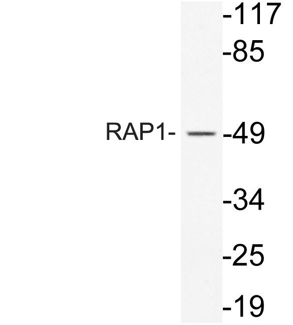
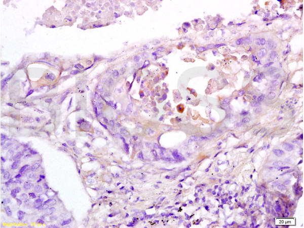
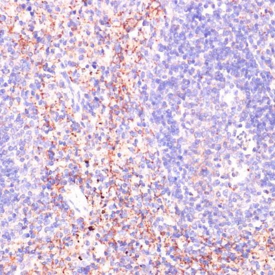
![IHC-P analysis of human kidney tissue using GTX31146 Rap1 antibody [1D2-1C64]. Right : Primary antibody Left : Negative control without primary antibody Antigen retrieval : 10mM sodium citrate (pH 6.0), microwaved for 8-15 min Dilution : 1:200](https://www.genetex.com/upload/website/prouct_img/normal/GTX31146/GTX31146_1345_IHC-P_w_23060722_488.webp)
Product group Antibodies
Rap1 antibody [1D2-1C64]GTX31146
ApplicationsImmunoFluorescence, ImmunoPrecipitation, Western Blot, ImmunoCytoChemistry, ImmunoHistoChemistry, ImmunoHistoChemistry Paraffin
ReactivityHuman, Mouse
TargetRAP1A
- SizePrice
Product group Antibodies
RAP1 antibodyGTX102770
ApplicationsWestern Blot
ReactivityHuman, Rat
TargetRAP1A
- SizePrice
Product group Antibodies
RAP1A antibodyGTX64554
ApplicationsWestern Blot, ImmunoHistoChemistry, ImmunoHistoChemistry Paraffin
ReactivityHuman, Mouse, Rat
TargetRAP1A
- SizePrice