

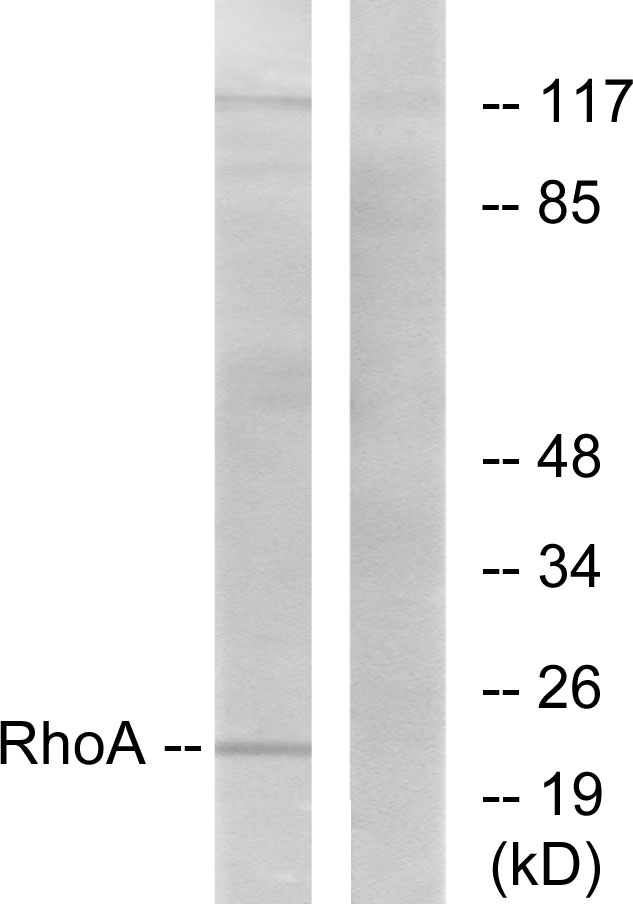
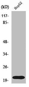
Western Blot analysis of K562, 231 and Hela cell, Human fetal brain tissue using RHOA Polyclonal Antibody at dilution of 1:400
Western Blot analysis of K562, 231 and Hela cell, Human fetal brain tissue using RHOA Polyclonal Antibody at dilution of 1:400
RHOA Polyclonal Antibody
E-AB-15491
Product group Antibodies
Overview
- SupplierElabscience
- Product NameRHOA Polyclonal Antibody
- Delivery Days Customer12
- Applications SupplierELISA WB
- CertificationResearch Use Only
- Concentration0.2mg/ml
- Scientific DescriptionRegulates a signal transduction pathway linking plasma membrane receptors to the assembly of focal adhesions and actin stress fibers. Involved in a microtubule-dependent signal that is required for the myosin contractile ring formation during cell cycle cytokinesis. Plays an essential role in cleavage furrow formation. Required for the apical junction formation of keratinocyte cell-cell adhesion. Serves as a target for the yopT cysteine peptidase from Yersinia pestis, vector of the plague, and Yersinia pseudotuberculosis, which causes gastrointestinal disorders. Stimulates PKN2 kinase activity. May be an activator of PLCE1. Activated by ARHGEF2, which promotes the exchange of GDP for GTP. Essential for the SPATA13-mediated regulation of cell migration and adhesion assembly and disassembly. The MEMO1-RHOA-DIAPH1 signaling pathway plays an important role in ERBB2-dependent stabilization of microtubules at the cell cortex. It controls the localization of APC and CLASP2 to the cell membrane, via the regulation of GSK3B activity. In turn, membrane-bound APC allows the localization of the MACF1 to the cell membrane, which is required for microtubule capture and stabilization.
- UNSPSC12352203
Related products
Product group Antibodies
Anti-RhoA Antibody144-61586
ApplicationsWestern Blot, ImmunoHistoChemistry
ReactivityHuman, Mouse, Rat
TargetRHOA
- SizePrice
![WB analysis of wide type (WT) and Rho knockout (KO) HeLa cells using GTX02825 RhoA + RhoB + RhoC antibody [GT1228]. Dilution : 1:1000 Loading : 25microg](https://www.genetex.com/upload/website/prouct_img/normal/GTX02825/CutImage_A19106_KO-WB_01_(1068999)_w_23053123_889.webp)
Product group Antibodies
ApplicationsWestern Blot
ReactivityHuman
TargetRHOA
- SizePrice
Product group Antibodies
Rhoa Polyclonal AntibodyCAC07100
ApplicationsELISA, ImmunoHistoChemistry
TargetRHOA
- SizePrice
Product group Antibodies
References
RhoA Polyclonal AntibodyBS-1180R
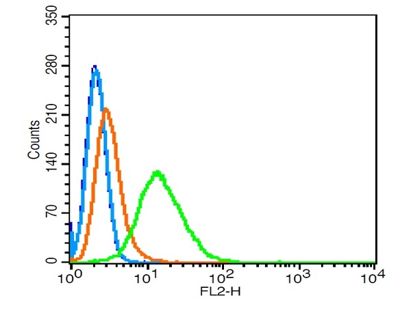
ApplicationsFlow Cytometry, ImmunoFluorescence, Western Blot, ELISA, ImmunoCytoChemistry, ImmunoHistoChemistry, ImmunoHistoChemistry Frozen, ImmunoHistoChemistry Paraffin
ReactivityCanine, Chicken, Human, Mouse, Rabbit, Rat, Zebra Fish
TargetRHOA
- SizePrice
Product group Antibodies
ApplicationsWestern Blot, ELISA, ImmunoCytoChemistry
ReactivityHuman
TargetRHOA
- SizePrice
Product group Antibodies
Anti-RhoA AntibodyA96128
ApplicationsWestern Blot, ELISA, ImmunoHistoChemistry
ReactivityHuman, Mouse, Rat
- SizePrice

Product group Antibodies
References
ApplicationsFlow Cytometry, ImmunoFluorescence, Western Blot, ImmunoCytoChemistry, ImmunoHistoChemistry
ReactivityHuman, Mouse, Rat
TargetRHOA
- SizePrice
Product group Antibodies
RHOA AntibodyCSB-PA003970
ApplicationsWestern Blot, ELISA, ImmunoHistoChemistry
ReactivityHuman, Mouse, Rat
TargetRHOA
- SizePrice