

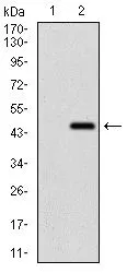
WB analysis of HEK293 (1) and Ring1 (AA: 79-263)-hIgGFc transfected HEK293 (2) cell lysate using GTX60753 Ring1 antibody [8C12F4].
![FACS analysis of HeLa cells using GTX60753 Ring1 antibody [8C12F4]. Green : Ring1 Red : negative control](https://www.genetex.com/upload/website/prouct_img/normal/GTX60753/GTX60753_20170912_FACS_w_23061123_129.webp "FACS analysis of HeLa cells using GTX60753 Ring1 antibody [8C12F4]. Green : Ring1 Red : negative control")
![ELISA analysis of antigen using GTX60753 Ring1 antibody [8C12F4].
Black : Control antigen 100ng
Purple : Antigen 10ng
Blue : Antigen 50ng
Red : Antigen 100ng](https://www.genetex.com/upload/website/prouct_img/normal/GTX60753/GTX60753_20170912_ELISA_w_23061123_308.webp "ELISA analysis of antigen using GTX60753 Ring1 antibody [8C12F4].
Black : Control antigen 100ng
Purple : Antigen 10ng
Blue : Antigen 50ng
Red : Antigen 100ng")
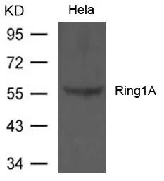
![WB analysis of MOLT-4 (1), LNCaP (2), HeLa (3), HEK-293 (4) and Jurkat (5) cell lysate using GTX60753 Ring1 antibody [8C12F4].](https://www.genetex.com/upload/website/prouct_img/normal/GTX60753/GTX60753_20170912_WB_1_w_23061123_345.webp "WB analysis of MOLT-4 (1), LNCaP (2), HeLa (3), HEK-293 (4) and Jurkat (5) cell lysate using GTX60753 Ring1 antibody [8C12F4].")
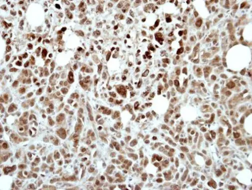
![IHC-P analysis of rectum cancer tissue using GTX60753 Ring1 antibody [8C12F4].](https://www.genetex.com/upload/website/prouct_img/normal/GTX60753/GTX60753_20170912_IHC-P_1_w_23061123_345.webp "IHC-P analysis of rectum cancer tissue using GTX60753 Ring1 antibody [8C12F4].")
![IHC-P analysis of cervical cancer tissue using GTX60753 Ring1 antibody [8C12F4].](https://www.genetex.com/upload/website/prouct_img/normal/GTX60753/GTX60753_20170912_IHC-P_w_23061123_233.webp "IHC-P analysis of cervical cancer tissue using GTX60753 Ring1 antibody [8C12F4].")
WB analysis of HEK293 (1) and Ring1 (AA: 79-263)-hIgGFc transfected HEK293 (2) cell lysate using GTX60753 Ring1 antibody [8C12F4].
Ring1 antibody [8C12F4]
GTX60753
ApplicationsFlow Cytometry, Western Blot, ELISA, ImmunoHistoChemistry, ImmunoHistoChemistry Paraffin
Product group Antibodies
ReactivityHuman
TargetRING1
Overview
- SupplierGeneTex
- Product NameRing1 antibody [8C12F4]
- Delivery Days Customer9
- Application Supplier NoteWB: 1/500 - 1/2000. IHC-P: 1/200 - 1/1000. FCM: 1/200 - 1/400. ELISA: 1/10000. *Optimal dilutions/concentrations should be determined by the researcher.Not tested in other applications.
- ApplicationsFlow Cytometry, Western Blot, ELISA, ImmunoHistoChemistry, ImmunoHistoChemistry Paraffin
- CertificationResearch Use Only
- ClonalityMonoclonal
- Clone ID8C12F4
- Concentration1 mg/ml
- ConjugateUnconjugated
- Gene ID6015
- Target nameRING1
- Target descriptionring finger protein 1
- Target synonymsRING1A, RNF1, E3 ubiquitin-protein ligase RING1, RING-type E3 ubiquitin transferase RING1, polycomb complex protein RING1, really interesting new gene 1 protein
- HostMouse
- IsotypeIgG1
- Protein IDQ06587
- Protein NameE3 ubiquitin-protein ligase RING1
- Scientific DescriptionThis gene belongs to the RING finger family, members of which encode proteins characterized by a RING domain, a zinc-binding motif related to the zinc finger domain. The gene product can bind DNA and can act as a transcriptional repressor. It is associated with the multimeric polycomb group protein complex. The gene product interacts with the polycomb group proteins BMI1, EDR1, and CBX4, and colocalizes with these proteins in large nuclear domains. It interacts with the CBX4 protein via its glycine-rich C-terminal domain. The gene maps to the HLA class II region, where it is contiguous with the RING finger genes FABGL and HKE4. [provided by RefSeq, Jul 2008]
- ReactivityHuman
- Storage Instruction-20°C or -80°C,2°C to 8°C
- UNSPSC41116161
Datasheet
Related products
Product group Antibodies
Anti-RING1 (N-term) Antibody102-21924
ApplicationsWestern Blot
TargetRING1
- SizePrice

Product group Antibodies
Anti-RING1 AntibodyA305946
ApplicationsWestern Blot, ImmunoHistoChemistry
ReactivityHuman, Mouse, Rat
- SizePrice

Product group Antibodies
Anti-RING1 Antibody Picoband(r)A01824-2-CARRIER-FREE
ApplicationsFlow Cytometry, ImmunoFluorescence, Western Blot, ELISA, ImmunoCytoChemistry
ReactivityHuman, Rat
TargetRING1
- SizePrice
Product group Antibodies
RING1 Recombinant Antibody, AbBy Fluor-405 ConjugatedBSM-62107R-BF405
ApplicationsImmunoFluorescence, Western Blot
ReactivityHuman
TargetRING1
- SizePrice

Product group Antibodies
RING1 AntibodyCSB-PA205793
ApplicationsWestern Blot, ELISA
ReactivityHuman, Mouse, Rat
TargetRING1
- SizePrice
Product group Antibodies

RING1 Antibody (aa150-200)LS-C290218
ApplicationsImmunoPrecipitation, Western Blot, ImmunoHistoChemistry
ReactivityHuman
TargetRING1
- SizePrice
Product group Antibodies
RING1 antibody [N1N3]GTX101671
ApplicationsWestern Blot, ImmunoHistoChemistry, ImmunoHistoChemistry Paraffin
ReactivityHuman
TargetRING1
- SizePrice

Product group Antibodies
Anti-RING1-25ulHPA008701
ApplicationsWestern Blot, ImmunoCytoChemistry, ImmunoHistoChemistry
ReactivityHuman
- SizePrice
Product group Antibodies
Ring1A antibodyGTX50803
ApplicationsWestern Blot
ReactivityHuman
TargetRING1
- SizePrice