

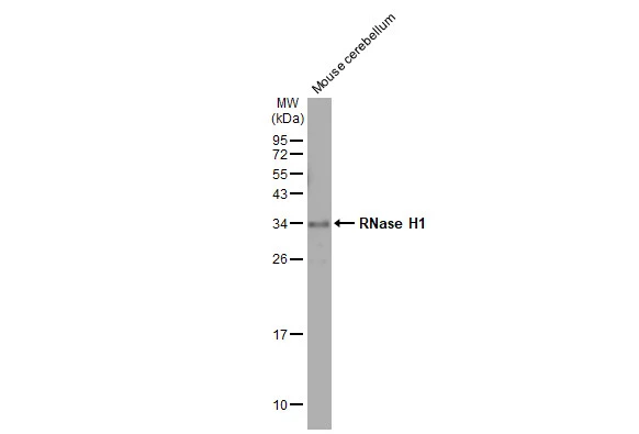
Mouse tissue extract (50 μg) was separated by 12% SDS-PAGE, and the membrane was blotted with RNase H1 antibody [HL2343] (GTX638546) diluted at 1:2000. The HRP-conjugated anti-rabbit IgG antibody (GTX213110-01) was used to detect the primary antibody.
![RNase H1 antibody [HL2343] detects RNase H1 protein at cytoplasm by immunohistochemical analysis. Sample: Paraffin-embedded human breast carcinoma. RNase H1 stained by RNase H1 antibody [HL2343] (GTX638546) diluted at 1:100. Antigen Retrieval: Citrate buffer, pH 6.0, 15 min](https://www.genetex.com/upload/website/prouct_img/normal/GTX638546/GTX638546_T-45026_20230512_IHC-P_23060622_825.webp "RNase H1 antibody [HL2343] detects RNase H1 protein at cytoplasm by immunohistochemical analysis. Sample: Paraffin-embedded human breast carcinoma. RNase H1 stained by RNase H1 antibody [HL2343] (GTX638546) diluted at 1:100. Antigen Retrieval: Citrate buffer, pH 6.0, 15 min")
![Whole cell extract (30 μg) was separated by 12% SDS-PAGE, and the membrane was blotted with RNase H1 antibody [HL2343] (GTX638546) diluted at 1:1000. The HRP-conjugated anti-rabbit IgG antibody (GTX213110-01) was used to detect the primary antibody.](https://www.genetex.com/upload/website/prouct_img/normal/GTX638546/GTX638546_T-45026_20230616_WB_D_23062019_596.webp "Whole cell extract (30 μg) was separated by 12% SDS-PAGE, and the membrane was blotted with RNase H1 antibody [HL2343] (GTX638546) diluted at 1:1000. The HRP-conjugated anti-rabbit IgG antibody (GTX213110-01) was used to detect the primary antibody.")
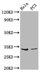
![Various whole cell extracts (30 μg) were separated by 12% SDS-PAGE, and the membrane was blotted with RNase H1 antibody [HL2343] (GTX638546) diluted at 1:1000. The HRP-conjugated anti-rabbit IgG antibody (GTX213110-01) was used to detect the primary antibody.](https://www.genetex.com/upload/website/prouct_img/normal/GTX638546/GTX638546_45110_20230721_WB_23072519_483.webp "Various whole cell extracts (30 μg) were separated by 12% SDS-PAGE, and the membrane was blotted with RNase H1 antibody [HL2343] (GTX638546) diluted at 1:1000. The HRP-conjugated anti-rabbit IgG antibody (GTX213110-01) was used to detect the primary antibody.")
![RNase H1 antibody [HL2343] detects RNase H1 protein by immunohistochemical analysis. Sample: Paraffin-embedded dog tissues. RNase H1 stained by RNase H1 antibody [HL2343] (GTX638546) diluted at 1:100. Antigen Retrieval: Citrate buffer, pH 6.0, 15 min](https://www.genetex.com/upload/website/prouct_img/normal/GTX638546/GTX638546_45110_20230829_IHC-P_multiple_Dog_23091319_152.webp "RNase H1 antibody [HL2343] detects RNase H1 protein by immunohistochemical analysis. Sample: Paraffin-embedded dog tissues. RNase H1 stained by RNase H1 antibody [HL2343] (GTX638546) diluted at 1:100. Antigen Retrieval: Citrate buffer, pH 6.0, 15 min")
Mouse tissue extract (50 μg) was separated by 12% SDS-PAGE, and the membrane was blotted with RNase H1 antibody [HL2343] (GTX638546) diluted at 1:2000. The HRP-conjugated anti-rabbit IgG antibody (GTX213110-01) was used to detect the primary antibody.
RNase H1 antibody [HL2343]
GTX638546
ApplicationsWestern Blot, ImmunoHistoChemistry, ImmunoHistoChemistry Paraffin
Product group Antibodies
ReactivityCanine, Human, Mouse
TargetRNASEH1
Overview
- SupplierGeneTex
- Product NameRNase H1 antibody [HL2343]
- Delivery Days Customer9
- Application Supplier NoteWB: 1:500-1:3000. *Optimal dilutions/concentrations should be determined by the researcher.Not tested in other applications.
- ApplicationsWestern Blot, ImmunoHistoChemistry, ImmunoHistoChemistry Paraffin
- CertificationResearch Use Only
- ClonalityMonoclonal
- Clone IDHL2343
- Concentration1 mg/ml
- ConjugateUnconjugated
- Gene ID246243
- Target nameRNASEH1
- Target descriptionribonuclease H1
- Target synonymsH1RNA, PEOB2, RNH1, ribonuclease H1, ribonuclease H type II
- HostRabbit
- IsotypeIgG
- Protein IDO60930
- Protein NameRibonuclease H1
- Scientific DescriptionThis gene encodes an endonuclease that specifically degrades the RNA of RNA-DNA hybrids and plays a key role in DNA replication and repair. Alternate in-frame start codon initiation results in the production of alternate isoforms that are directed to the mitochondria or to the nucleus. The production of the mitochondrial isoform is modulated by an upstream open reading frame (uORF). Mutations in this gene have been found in individuals with progressive external ophthalmoplegia with mitochondrial DNA deletions, autosomal recessive 2. Alternative splicing results in additional coding and non-coding transcript variants. Pseudogenes of this gene have been defined on chromosomes 2 and 17. [provided by RefSeq, Jul 2017]
- ReactivityCanine, Human, Mouse
- Storage Instruction-20°C or -80°C,2°C to 8°C
- UNSPSC41116161
Datasheet
Related products
Product group Antibodies
RNASEH1 AntibodyCSB-PA019801LA01HU
ApplicationsWestern Blot, ELISA, ImmunoHistoChemistry
ReactivityHuman
TargetRNASEH1
- SizePrice
Product group Antibodies
Rnaseh1 Polyclonal AntibodyCAC11443
ApplicationsWestern Blot, ELISA, ImmunoHistoChemistry
TargetRNASEH1
- SizePrice

Product group Antibodies
Anti-RNASEH1 Antibody Picoband(r)A06342-1-CARRIER-FREE
ApplicationsFlow Cytometry, ImmunoFluorescence, Western Blot, ELISA, ImmunoCytoChemistry
ReactivityHuman, Mouse
TargetRNASEH1
- SizePrice

Product group Antibodies
Anti-RNASEH1 AntibodyHPA034817
ApplicationsWestern Blot, ImmunoCytoChemistry
ReactivityHuman
TargetRNASEH1
- SizePrice
Product group Antibodies

RNASEH1 AntibodyLS-C410650
ApplicationsImmunoPrecipitation, Western Blot
ReactivityHuman, Mouse, Rat
TargetRNASEH1
- SizePrice
![Various whole cell extracts (30 μg) were separated by 12% SDS-PAGE, and the membrane was blotted with RNase H1 antibody [N2C3] (GTX117624) diluted at 1:1000. The HRP-conjugated anti-rabbit IgG antibody (GTX213110-01) was used to detect the primary antibody.](https://www.genetex.com/upload/website/prouct_img/normal/GTX117624/GTX117624_44980_20230317_WB_23032819_551.webp)
Product group Antibodies
RNase H1 antibody [N2C3]GTX117624
ApplicationsWestern Blot, ImmunoHistoChemistry, ImmunoHistoChemistry Paraffin
ReactivityHuman, Zebra Fish
TargetRNASEH1
- SizePrice