

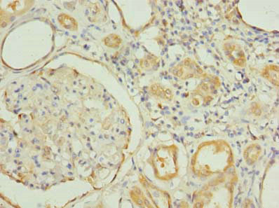
Immunohistochemistry of paraffin-embedded human kidney tissue using CSB-PA856927LA01HU at dilution of 1:100
")
Immunohistochemistry of paraffin-embedded human kidney tissue using CSB-PA856927LA01HU at dilution of 1:100
RNF31 Antibody
CSB-PA856927LA01HU
ApplicationsImmunoFluorescence, ELISA, ImmunoHistoChemistry
Product group Antibodies
ReactivityHuman
TargetRNF31
Overview
- SupplierCusabio
- Product NameRNF31 Antibody
- Delivery Days Customer20
- ApplicationsImmunoFluorescence, ELISA, ImmunoHistoChemistry
- CertificationResearch Use Only
- ClonalityPolyclonal
- ConjugateUnconjugated
- Gene ID55072
- Target nameRNF31
- Target descriptionring finger protein 31
- Target synonymsHOIP, IMD115, Paul, ZIBRA, E3 ubiquitin-protein ligase RNF31, HOIL-1-interacting protein, RING-type E3 ubiquitin transferase RNF31, zinc in-between-RING-finger ubiquitin-associated domain protein
- HostRabbit
- IsotypeIgG
- Protein IDQ96EP0
- Protein NameE3 ubiquitin-protein ligase RNF31
- Scientific DescriptionE3 ubiquitin-protein ligase component of the LUBAC complex which conjugates linear (Met-1-linked) polyubiquitin chains to substrates and plays a key role in NF-kappa-B activation and regulation of inflammation. LUBAC conjugates linear polyubiquitin to IKBKG and RIPK1 and is involved in activation of the canonical NF-kappa-B and the JNK signaling pathways. Linear ubiquitination mediated by the LUBAC complex interferes with TNF-induced cell death and thereby prevents inflammation. LUBAC is proposed to be recruited to the TNF-R1 signaling complex (TNF-RSC) following polyubiquitination of TNF-RSC components by BIRC2 and/or BIRC3 and to conjugate linear polyubiquitin to IKBKG and possibly other components contributing to the stability of the complex. Together with FAM105B/otulin, the LUBAC complex regulates the canonical Wnt signaling during angiogenesis. Binds polyubiquitin of different linkage types.
- ReactivityHuman
- Storage Instruction-20°C or -80°C
- UNSPSC41116161
Related products

Product group Antibodies
Anti-RNF31/HOIP Antibody Picoband(r)A04457-3-CARRIER-FREE
ApplicationsWestern Blot, ELISA
ReactivityHuman, Mouse, Rat
TargetRNF31
- SizePrice
Product group Antibodies
Anti-RNF31 Antibody144-08227
ApplicationsWestern Blot
ReactivityHuman
TargetRNF31
- SizePrice

Product group Antibodies

RNF31 AntibodyLS-C749150
ApplicationsWestern Blot
ReactivityHuman, Mouse
TargetRNF31
- SizePrice
Product group Antibodies
RNF31 Polyclonal AntibodyBS-9241R
ApplicationsFlow Cytometry, ImmunoFluorescence, ImmunoCytoChemistry, ImmunoHistoChemistry, ImmunoHistoChemistry Frozen, ImmunoHistoChemistry Paraffin
ReactivityHuman, Mouse, Rat
TargetRNF31
- SizePrice
Product group Antibodies

ApplicationsWestern Blot, ELISA
ReactivityCanine, Human, Porcine
TargetRNF31
- SizePrice

Product group Antibodies
Anti-RNF31 AntibodyHPA048745
ApplicationsImmunoCytoChemistry, ImmunoHistoChemistry
ReactivityHuman
TargetRNF31
- SizePrice
![RNF31 antibody [HL2295] detects RNF31 protein by immunohistochemical analysis. Sample: Paraffin-embedded rat tissues. RNF31 stained by RNF31 antibody [HL2295] (GTX638350) diluted at 1:100. Antigen Retrieval: Citrate buffer, pH 6.0, 15 min](https://www.genetex.com/upload/website/prouct_img/normal/GTX638350/GTX638350_T-44984_20230331_IHC-P_multiple_R_23041023_680.webp)
Product group Antibodies
RNF31 antibody [HL2295]GTX638350
ApplicationsWestern Blot, ImmunoHistoChemistry, ImmunoHistoChemistry Paraffin
ReactivityHuman, Mouse, Rat
TargetRNF31
- SizePrice