

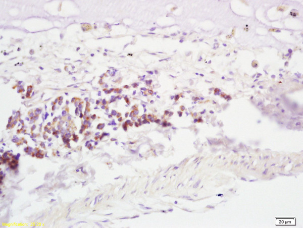
Formalin-fixed and paraffin embedded human lung carcinoma labeled with Anti-RNF43 Polyclonal Antibody, Unconjugated(bs-7007R) at 1:200, followed by conjugation to the secondary antibody and DAB staining
 followed by conjugation to the secondary antibody and DAB staining")
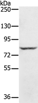
 at 1:1000 dilution and 4˚C overnight incubation. Followed by conjugated secondary antibody incubation at 1:20000 for 60 min at 37˚C.")
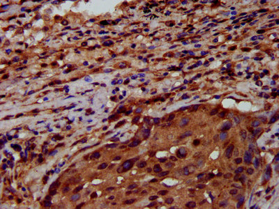
Formalin-fixed and paraffin embedded human lung carcinoma labeled with Anti-RNF43 Polyclonal Antibody, Unconjugated(bs-7007R) at 1:200, followed by conjugation to the secondary antibody and DAB staining
RNF43 Polyclonal Antibody
BS-7007R
ApplicationsImmunoFluorescence, Western Blot, ELISA, ImmunoCytoChemistry, ImmunoHistoChemistry, ImmunoHistoChemistry Frozen, ImmunoHistoChemistry Paraffin
Product group Antibodies
ReactivityBovine, Canine, Equine, Human, Mouse, Porcine, Rat
TargetRNF43
Overview
- SupplierBioss
- Product NameRNF43 Polyclonal Antibody
- Delivery Days Customer16
- ApplicationsImmunoFluorescence, Western Blot, ELISA, ImmunoCytoChemistry, ImmunoHistoChemistry, ImmunoHistoChemistry Frozen, ImmunoHistoChemistry Paraffin
- Applications SupplierWB(1:300-5000), ELISA(1:500-1000), IHC-P(1:200-400), IHC-F(1:100-500), IF(IHC-P)(1:50-200), IF(IHC-F)(1:50-200), IF(ICC)(1:50-200)
- CertificationResearch Use Only
- ClonalityPolyclonal
- Concentration1 ug/ul
- ConjugateUnconjugated
- Gene ID54894
- Target nameRNF43
- Target descriptionring finger protein 43
- Target synonymsRNF124, SSPCS, URCC, E3 ubiquitin-protein ligase RNF43, RING-type E3 ubiquitin transferase RNF43
- HostRabbit
- IsotypeIgG
- Protein IDQ68DV7
- Protein NameE3 ubiquitin-protein ligase RNF43
- ReactivityBovine, Canine, Equine, Human, Mouse, Porcine, Rat
- Storage Instruction-20°C
- UNSPSC41116161
Datasheet
Related products
Product group Antibodies
Anti-RNF43 (C-term) Antibody102-21400
ApplicationsWestern Blot, ImmunoHistoChemistry, ImmunoHistoChemistry Paraffin
TargetRNF43
- SizePrice
Product group Antibodies
ApplicationsOther Application
TargetRNF43
- SizePrice
Product group Antibodies

RNF43 AntibodyLS-C672876
ApplicationsELISA, ImmunoHistoChemistry, ImmunoHistoChemistry Paraffin
ReactivityHuman
TargetRNF43
- SizePrice

Product group Antibodies
Anti-RNF43 Antibody Picoband(r)A01694-1-CARRIER-FREE
ApplicationsWestern Blot, ELISA
ReactivityHuman
TargetRNF43
- SizePrice
Product group Antibodies
RNF43 Polyclonal AntibodyCAC13189
ApplicationsELISA, ImmunoHistoChemistry
TargetRNF43
- SizePrice
Product group Antibodies
RNF43 AntibodyCSB-PA019892LA01HU
ApplicationsELISA, ImmunoHistoChemistry
ReactivityHuman
TargetRNF43
- SizePrice
Product group Antibodies
RNF43 antibodyGTX132671
ApplicationsWestern Blot
ReactivityHuman, Mouse
TargetRNF43
- SizePrice

Product group Antibodies
Anti-RNF43 AntibodyHPA008079
ApplicationsWestern Blot, ImmunoHistoChemistry
ReactivityHuman
TargetRNF43
- SizePrice