

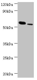
Western blot All lanes: RNF8 antibody at 11microg/ml Lane 1: Hela whole cell lysate Lane 2: 293T whole cell lysate Secondary Goat polyclonal to rabbit IgG at 1/10000 dilution Predicted band size: 56, 11, 51 kDa Observed band size: 56 kDa

Western blot All lanes: RNF8 antibody at 11microg/ml Lane 1: Hela whole cell lysate Lane 2: 293T whole cell lysate Secondary Goat polyclonal to rabbit IgG at 1/10000 dilution Predicted band size: 56, 11, 51 kDa Observed band size: 56 kDa
RNF8 Antibody
CSB-PA019898ESR2HU
ApplicationsWestern Blot, ELISA, ImmunoHistoChemistry
Product group Antibodies
ReactivityHuman
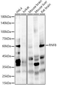
TargetRNF8
Overview
- SupplierCusabio
- Product NameRNF8 Antibody
- Delivery Days Customer20
- ApplicationsWestern Blot, ELISA, ImmunoHistoChemistry
- CertificationResearch Use Only
- ClonalityPolyclonal
- ConjugateUnconjugated
- Gene ID9025
- Target nameRNF8
- Target descriptionring finger protein 8
- Target synonymshRNF8, E3 ubiquitin-protein ligase RNF8, C3HC4-type zinc finger protein, RING-type E3 ubiquitin transferase RNF8, UBC13/UEV-interacting ring finger protein, ring finger protein (C3HC4 type) 8, ring finger protein 8, E3 ubiquitin protein ligase
- HostRabbit
- IsotypeIgG
- Protein IDO76064
- Protein NameE3 ubiquitin-protein ligase RNF8
- Scientific DescriptionE3 ubiquitin-protein ligase that plays a key role in DNA damage signaling via 2 distinct roles: by mediating the Lys-63-linked ubiquitination of histones H2A and H2AX and promoting the recruitment of DNA repair proteins at double-strand breaks (DSBs) sites, and by catalyzing Lys-48-linked ubiquitination to remove target proteins from DNA damage sites. Following DNA DSBs, it is recruited to the sites of damage by ATM-phosphorylated MDC1 and catalyzes the Lys-63-linked ubiquitination of histones H2A and H2AX, thereby promoting the formation of TP53BP1 and BRCA1 ionizing radiation-induced foci (IRIF). Also controls the recruitment of UIMC1-BRCC3 (RAP80-BRCC36) and PAXIP1/PTIP to DNA damage sites. Also recruited at DNA interstrand cross-links (ICLs) sites and catalyzes Lys-63-linked ubiquitination of histones H2A and H2AX, leading to recruitment of FAAP20/C1orf86 and Fanconi anemia (FA) complex, followed by interstrand cross-link repair. H2A ubiquitination also mediates the ATM-dependent transcriptional silencing at regions flanking DSBs in cis, a mechanism to avoid collision between transcription and repair intermediates. Promotes the formation of Lys-63-linked polyubiquitin chains via interactions with the specific ubiquitin-conjugating UBE2N/UBC13 and ubiquitinates non-histone substrates such as PCNA. Substrates that are polyubiquitinated at Lys-63 are usually not targeted for degradation. Also catalyzes the formation of Lys-48-linked polyubiquitin chains via interaction with the ubiquitin-conjugating UBE2L6/UBCH8, leading to degradation of substrate proteins such as CHEK2, JMJD2A/KDM4A and KU80/XRCC5: it is still unclear how the preference toward Lys-48- versus Lys-63-linked ubiquitination is regulated but it could be due to RNF8 ability to interact with specific E2 specific ligases. For instance, interaction with phosphorylated HERC2 promotes the association between RNF8 and UBE2N/UBC13 and favors the specific formation of Lys-63-linked ubiquitin chains. Promotes non-homologous end joining (NHEJ) by promoting the Lys-48-linked ubiquitination and degradation the of KU80/XRCC5. Following DNA damage, mediates the ubiquitination and degradation of JMJD2A/KDM4A in collaboration with RNF168, leading to unmask H4K20me2 mark and promote the recruitment of TP53BP1 at DNA damage sites. In addition to its function in damage signaling, also plays a role in higher-order chromatin structure by mediating extensive chromatin decondensation. Involved in the activation of ATM by promoting histone H2B ubiquitination, which indirectly triggers histone H4 Lys-16 acetylation (H4K16ac), establishing a chromatin environment that promotes efficient activation of ATM kinase. Required in the testis, where it plays a role in the replacement of histones during spermatogenesis. At uncapped telomeres, promotes the joining of deprotected chromosome ends by inducing H2A ubiquitination and TP53BP1 recruitment, suggesting that it may enhance cancer development by aggravating telomere-induced genome instability in case of telomeric crisis. Promotes the assembly of RAD51 at DNA DSBs in the absence of BRCA1 and TP53BP1 Also involved in class switch recombination in immune system, via its role in regulation of DSBs repair. May be required for proper exit from mitosis after spindle checkpoint activation and may regulate cytokinesis. May play a role in the regulation of RXRA-mediated transcriptional activity. Not involved in RXRA ubiquitination by UBE2E2.
- ReactivityHuman
- Storage Instruction-20°C or -80°C
- UNSPSC41116161
Related products
Product group Antibodies
Anti-RNF8 AntibodyA10107
ApplicationsWestern Blot
ReactivityHuman, Mouse, Rat
- SizePrice

Product group Antibodies
Anti-RNF8 Antibody Picoband(r)A00707-2-CARRIER-FREE
ApplicationsFlow Cytometry, Western Blot, ELISA
ReactivityHuman
TargetRNF8
- SizePrice
Product group Antibodies

RNF8 AntibodyLS-C749096
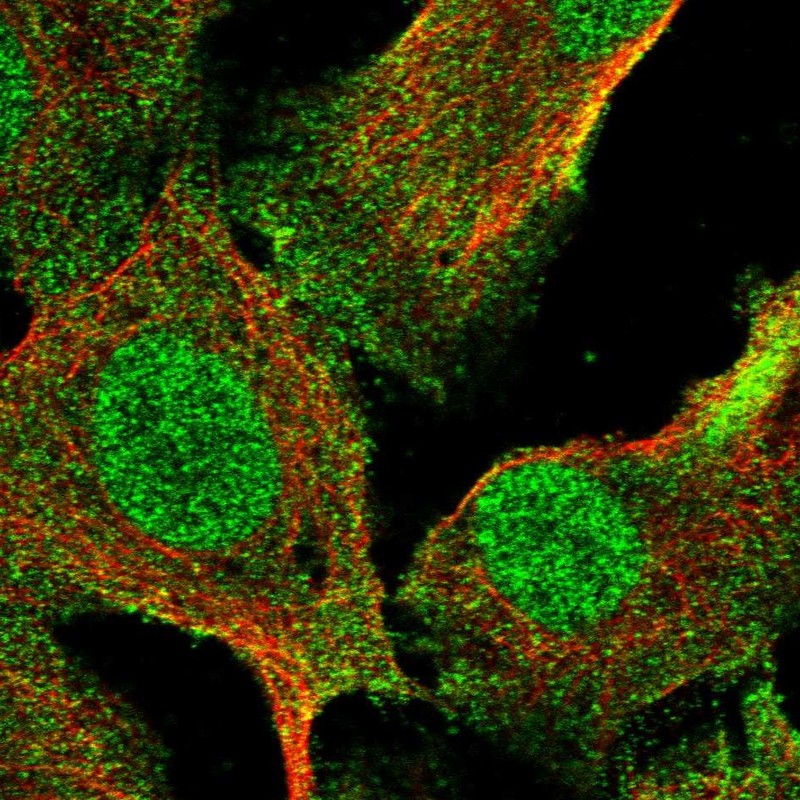
ApplicationsImmunoFluorescence, Western Blot
ReactivityHuman
TargetRNF8
- SizePrice
Product group Antibodies
RNF8 Recombinant Antibody, AbBy Fluor-488 ConjugatedBSM-61658R-BF488
ApplicationsWestern Blot
ReactivityHuman, Rat
TargetRNF8
- SizePrice
Product group Antibodies

Goat anti-RNF8EB05843
ApplicationsFlow Cytometry, Western Blot, ELISA
ReactivityHuman
TargetRNF8
- SizePrice
Product group Antibodies
Anti-RNF8 AntibodyHPA064925
ApplicationsImmunoCytoChemistry
ReactivityHuman
TargetRNF8
- SizePrice
Product group Antibodies
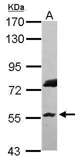
RNF8 antibody [N3C3]GTX115176
ApplicationsWestern Blot
ReactivityHuman
TargetRNF8
- SizePrice