

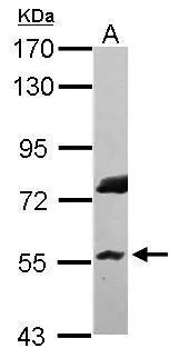
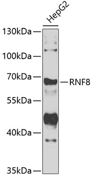
Sample (30 ug of whole cell lysate) A: HepG2 7.5% SDS PAGE GTX115176 diluted at 1:5000
Sample (30 ug of whole cell lysate) A: HepG2 7.5% SDS PAGE GTX115176 diluted at 1:5000
RNF8 antibody [N3C3]
GTX115176
ApplicationsWestern Blot
Product group Antibodies
ReactivityHuman
TargetRNF8
Overview
- SupplierGeneTex
- Product NameRNF8 antibody [N3C3]
- Delivery Days Customer9
- Application Supplier NoteWB: 1:1000-1:10000. *Optimal dilutions/concentrations should be determined by the researcher.Not tested in other applications.
- ApplicationsWestern Blot
- CertificationResearch Use Only
- ClonalityPolyclonal
- Concentration0.86 mg/ml
- ConjugateUnconjugated
- Gene ID9025
- Target nameRNF8
- Target descriptionring finger protein 8
- Target synonymshRNF8, E3 ubiquitin-protein ligase RNF8, C3HC4-type zinc finger protein, RING-type E3 ubiquitin transferase RNF8, UBC13/UEV-interacting ring finger protein, ring finger protein (C3HC4 type) 8, ring finger protein 8, E3 ubiquitin protein ligase
- HostRabbit
- IsotypeIgG
- Protein IDO76064
- Protein NameE3 ubiquitin-protein ligase RNF8
- Scientific DescriptionThe protein encoded by this gene contains a RING finger motif and a FHA domain. This protein has been shown to interact with several class II ubiquitin-conjugating enzymes (E2), including UBE2E1/UBCH6, UBE2E2, and UBE2E3, and may act as an ubiquitin ligase (E3) in the ubiquitination of certain nuclear proteins. Alternatively spliced transcript variants encoding distinct isoforms have been reported. [provided by RefSeq]
- ReactivityHuman
- Storage Instruction-20°C or -80°C,2°C to 8°C
- UNSPSC41116161
Datasheet
Related products
Product group Antibodies
Anti-RNF8 AntibodyA10107
ApplicationsWestern Blot
ReactivityHuman, Mouse, Rat
- SizePrice

Product group Antibodies
Anti-RNF8 Antibody Picoband(r)A00707-2-CARRIER-FREE
ApplicationsFlow Cytometry, Western Blot, ELISA
ReactivityHuman
TargetRNF8
- SizePrice
Product group Antibodies

RNF8 AntibodyLS-C749096
ApplicationsImmunoFluorescence, Western Blot
ReactivityHuman
TargetRNF8
- SizePrice
Product group Antibodies
RNF8 Recombinant Antibody, AbBy Fluor-488 ConjugatedBSM-61658R-BF488
ApplicationsWestern Blot
ReactivityHuman, Rat
TargetRNF8
- SizePrice
Product group Antibodies

Goat anti-RNF8EB05843
ApplicationsFlow Cytometry, Western Blot, ELISA
ReactivityHuman
TargetRNF8
- SizePrice
Product group Antibodies
RNF8 AntibodyCSB-PA019898ESR2HU
ApplicationsWestern Blot, ELISA, ImmunoHistoChemistry
ReactivityHuman
TargetRNF8
- SizePrice
Product group Antibodies
RNF8 antibody, N-termGTX15850
ApplicationsWestern Blot
ReactivityHuman
TargetRNF8
- SizePrice
Product group Antibodies
Anti-RNF8 AntibodyHPA064925
ApplicationsImmunoCytoChemistry
ReactivityHuman
TargetRNF8
- SizePrice
Product group Antibodies
RNF8 antibodyGTX55783
ApplicationsWestern Blot
ReactivityHuman
TargetRNF8
- SizePrice