Bio-Connect
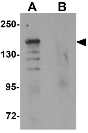
WB analysis of 293 cell lysate in (A) the absence and (B) the presence of blocking peptide using GTX31836 ROCK1 antibody. Working concentration : 1 microg/ml

ROCK1 antibody

GTX31836
GeneTex
ApplicationsImmunoFluorescence, Western Blot, ELISA, ImmunoCytoChemistry
Product group Antibodies
TargetROCK1
Sign in to order and to see your custom pricing.
Large volume orders?
Order with a bulk request

Overview

  • Supplier
    GeneTex
  • Product Name
    ROCK1 antibody
  • Delivery Days Customer
    9
  • Application Supplier Note
    WB: 1 microg/mL. ICC/IF: 10 microg/mL. *Optimal dilutions/concentrations should be determined by the researcher.Not tested in other applications.
  • Applications
    ImmunoFluorescence, Western Blot, ELISA, ImmunoCytoChemistry
  • Certification
    Research Use Only
  • Clonality
    Polyclonal
  • Concentration
    1 mg/ml
  • Conjugate
    Unconjugated
  • Gene ID6093
  • Target name
    ROCK1
  • Target description
    Rho associated coiled-coil containing protein kinase 1
  • Target synonyms
    p160 ROCK-1; P160ROCK; renal carcinoma antigen NY-REN-35; rho-associated protein kinase 1; ROCK-I
  • Host
    Rabbit
  • Isotype
    IgG
  • Protein IDQ13464
  • Protein Name
    Rho-associated protein kinase 1
  • Scientific Description
    This gene encodes a protein serine/threonine kinase that is activated when bound to the GTP-bound form of Rho. The small GTPase Rho regulates formation of focal adhesions and stress fibers of fibroblasts, as well as adhesion and aggregation of platelets and lymphocytes by shuttling between the inactive GDP-bound form and the active GTP-bound form. Rho is also essential in cytokinesis and plays a role in transcriptional activation by serum response factor. This protein, a downstream effector of Rho, phosphorylates and activates LIM kinase, which in turn, phosphorylates cofilin, inhibiting its actin-depolymerizing activity. [provided by RefSeq, Jul 2008]
  • Storage Instruction
    -20°C or -80°C,2°C to 8°C
  • UNSPSC
    12352203

Related products

Product group Antibodies
Biomatik
ApplicationsImmunoFluorescence, Western Blot, ELISA, ImmunoHistoChemistry
TargetROCK1
  • SizePrice
Product group Antibodies
Assay Genie
TargetROCK1
  • SizePrice
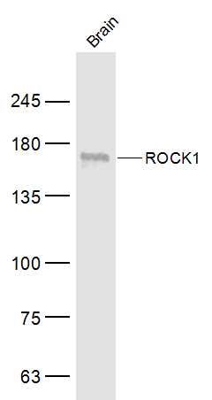
Mouse brain lysates probed with ROCK1 Polyclonal Antibody, Unconjugated (bs-1166R) at 1:500 dilution and 4˚C overnight incubation. Followed by conjugated secondary antibody incubation at 1:10000 for 60 min at 37˚C.
Product group Antibodies
References
Bioss Antibodies
ApplicationsImmunoFluorescence, Western Blot, ELISA, ImmunoCytoChemistry, ImmunoHistoChemistry, ImmunoHistoChemistry Frozen, ImmunoHistoChemistry Paraffin
TargetROCK1
  • SizePrice
Product group Antibodies
Bethyl Laboratories
ApplicationsImmunoPrecipitation
TargetROCK1
  • SizePrice
Anti-Rock-1 Antibody
Product group Antibodies
Antibodies.com
ApplicationsWestern Blot, ELISA, ImmunoHistoChemistry
  • SizePrice
Product group Antibodies
RayBiotech
ApplicationsImmunoPrecipitation, Western Blot, ImmunoHistoChemistry
TargetROCK1
  • SizePrice
IP analysis of 293T cell lysate using GTX32843 ROCK1 antibody. Antibody amount : 3microg / 200microg lysate Dilution : 1:1000
Product group Antibodies
GeneTex
ApplicationsImmunoPrecipitation, Western Blot, ImmunoHistoChemistry, ImmunoHistoChemistry Paraffin
TargetROCK1
  • SizePrice
Product group Antibodies
MedChem Express
TargetROCK1
  • SizePrice
Figure 1. Western blot analysis of ROCK1 using anti-ROCK1 antibody (A00722-4). Electrophoresis was performed on a 5-20% SDS-PAGE gel at 70V (Stacking gel) / 90V (Resolving gel) for 2-3 hours. The sample well of each lane was loaded with 30 ug of sample under reducing conditions. Lane 1: human Hela whole cell lysates, Lane 2: monkey COS-7 whole cell lysates, Lane 3: rat liver tissue lysates, Lane 4: rat C6 whole cell lysates, Lane 5: mouse liver tissue lysates, Lane 6: mouse NIH/3T3 whole cell lysates. After electrophoresis, proteins were transferred to a nitrocellulose membrane at 150 mA for 50-90 minutes. Blocked the membrane with 5% non-fat milk/TBS for 1.5 hour at RT. The membrane was incubated with rabbit anti-ROCK1 antigen affinity purified polyclonal antibody (Catalog # A00722-4) at 0.5 microg/mL overnight at 4°C, then washed with TBS-0.1%Tween 3 times with 5 minutes each and probed with a goat anti-rabbit IgG-HRP secondary antibody at a dilution of 1:5000 for 1.5 hour at RT. The signal is developed using an Enhanced Chemiluminescent detection (ECL) kit (Catalog # EK1002) with Tanon 5200 system. A specific band was detected for ROCK1 at approximately 160 kDa. The expected band size for ROCK1 is at 158 kDa.
Product group Antibodies
Boster Bio
Anti-ROCK1 Antibody Picoband(r)A00722-4-CARRIER-FREE
ApplicationsFlow Cytometry, Western Blot, ELISA
TargetROCK1
  • SizePrice
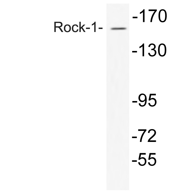
Western Blot analysis of COS7 cells using Rock-1 Polyclonal Antibody
Product group Antibodies
Cusabio
ROCK1 AntibodyCSB-PA004017
ApplicationsWestern Blot, ELISA, ImmunoHistoChemistry
ReactivityHuman, Monkey, Mouse, Rat
TargetROCK1
  • SizePrice