

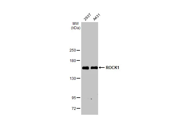
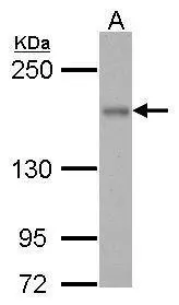
Various whole cell extracts (30 μg) were separated by 5% SDS-PAGE, and the membrane was blotted with ROCK1 antibody [N1N2], N-term (GTX113266) diluted at 1:1000. The HRP-conjugated anti-rabbit IgG antibody (GTX213110-01) was used to detect the primary antibody.
![ROCK1 antibody [N1N2], N-term detects ROCK1 protein by western blot analysis. A. 30 μg NIH-3T3 whole cell extract B. 30 μg C2Cl2 whole cell extract 5% SDS-PAGE ROCK1 antibody [N1N2], N-term (GTX113266) dilution: 1:1000 The HRP-conjugated anti-rabbit IgG antibody (GTX213110-01) was used to detect the primary antibody.](https://www.genetex.com/upload/website/prouct_img/normal/GTX113266/GTX113266_40128_WB_M_w_23060501_196.webp "ROCK1 antibody [N1N2], N-term detects ROCK1 protein by western blot analysis. A. 30 μg NIH-3T3 whole cell extract B. 30 μg C2Cl2 whole cell extract 5% SDS-PAGE ROCK1 antibody [N1N2], N-term (GTX113266) dilution: 1:1000 The HRP-conjugated anti-rabbit IgG antibody (GTX213110-01) was used to detect the primary antibody.")
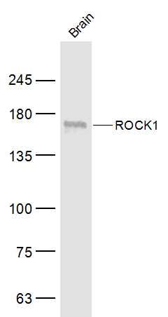
![ROCK1 antibody [N1N2], N-term detects ROCK1 protein by western blot analysis. A. 50 μg rat brain lysate/extract 5% SDS-PAGE ROCK1 antibody [N1N2], N-term (GTX113266) dilution: 1:500 The HRP-conjugated anti-rabbit IgG antibody (GTX213110-01) was used to detect the primary antibody.](https://www.genetex.com/upload/website/prouct_img/normal/GTX113266/GTX113266_40128_WB_R_brain_w_23060501_879.webp "ROCK1 antibody [N1N2], N-term detects ROCK1 protein by western blot analysis. A. 50 μg rat brain lysate/extract 5% SDS-PAGE ROCK1 antibody [N1N2], N-term (GTX113266) dilution: 1:500 The HRP-conjugated anti-rabbit IgG antibody (GTX213110-01) was used to detect the primary antibody.")
![ROCK1 antibody [N1N2], N-term detects ROCK1 protein at cytoplasm on human breast carcinoma by immunohistochemical analysis. Sample: Paraffin-embedded human breast carcinoma. ROCK1 antibody [N1N2], N-term (GTX113266) diluted at 1:500.
Antigen Retrieval: Trilogy? (EDTA based, pH 8.0) buffer, 15min](https://www.genetex.com/upload/website/prouct_img/normal/GTX113266/GTX113266_40128_20141219_IHC_w_23060501_407.webp "ROCK1 antibody [N1N2], N-term detects ROCK1 protein at cytoplasm on human breast carcinoma by immunohistochemical analysis. Sample: Paraffin-embedded human breast carcinoma. ROCK1 antibody [N1N2], N-term (GTX113266) diluted at 1:500.
Antigen Retrieval: Trilogy? (EDTA based, pH 8.0) buffer, 15min")
![ROCK1 antibody [N1N2], N-term detects ROCK1 protein at nucleus and Golgi apparatus by immunofluorescent analysis. Sample: HeLa cells were fixed in 4% paraformaldehyde at RT for 15 min. Green: ROCK1 stained by ROCK1 antibody [N1N2], N-term (GTX113266) diluted at 1:500. Red: alpha Tubulin, a cytoskeleton marker, stained by alpha Tubulin antibody [GT114] (GTX628802) diluted at 1:1000. Blue: Fluoroshield with DAPI (GTX30920).](https://www.genetex.com/upload/website/prouct_img/normal/GTX113266/GTX113266_44560_20220429_ICC_IF_w_23060501_312.webp "ROCK1 antibody [N1N2], N-term detects ROCK1 protein at nucleus and Golgi apparatus by immunofluorescent analysis. Sample: HeLa cells were fixed in 4% paraformaldehyde at RT for 15 min. Green: ROCK1 stained by ROCK1 antibody [N1N2], N-term (GTX113266) diluted at 1:500. Red: alpha Tubulin, a cytoskeleton marker, stained by alpha Tubulin antibody [GT114] (GTX628802) diluted at 1:1000. Blue: Fluoroshield with DAPI (GTX30920).")
![Wild-type (WT) and ROCK1 knockout (KO) HeLa cell extracts (30 μg) were separated by 5% SDS-PAGE, and the membrane was blotted with ROCK1 antibody [N1N2], N-term (GTX113266) diluted at 1:1000. The HRP-conjugated anti-rabbit IgG antibody (GTX213110-01) was used to detect the primary antibody.](https://www.genetex.com/upload/website/prouct_img/normal/GTX113266/GTX113266_43103_20190215_WB_KO_watermark_w_23060501_220.webp "Wild-type (WT) and ROCK1 knockout (KO) HeLa cell extracts (30 μg) were separated by 5% SDS-PAGE, and the membrane was blotted with ROCK1 antibody [N1N2], N-term (GTX113266) diluted at 1:1000. The HRP-conjugated anti-rabbit IgG antibody (GTX213110-01) was used to detect the primary antibody.")
![Non-transfected (–) and transfected (+) HeLa whole cell extracts (30 μg) were separated by 5% SDS-PAGE, and the membrane was blotted with ROCK1 antibody [N1N2], N-term (GTX113266) diluted at 1:1000. The HRP-conjugated anti-rabbit IgG antibody (GTX213110-01) was used to detect the primary antibody.](https://www.genetex.com/upload/website/prouct_img/normal/GTX113266/GTX113266_42256_20160728_WB_shRNA_watermark_w_23060501_783.webp "Non-transfected (–) and transfected (+) HeLa whole cell extracts (30 μg) were separated by 5% SDS-PAGE, and the membrane was blotted with ROCK1 antibody [N1N2], N-term (GTX113266) diluted at 1:1000. The HRP-conjugated anti-rabbit IgG antibody (GTX213110-01) was used to detect the primary antibody.")
![ROCK1 antibody [N1N2], N-term detects ROCK1 protein at cytoplasm by immunofluorescent analysis. Sample: HeLa cells were fixed in 4% paraformaldehyde at RT for 15 min. Green: ROCK1 protein stained by ROCK1 antibody [N1N2], N-term (GTX113266) diluted at 1:500. Red: alpha Tubulin, a cytoskeleton marker, stained by alpha Tubulin antibody [GT114] (GTX628802) diluted at 1:500. Blue: Hoechst 33342 staining.](https://www.genetex.com/upload/website/prouct_img/normal/GTX113266/GTX113266_40128_20150410_IFA_w_23060501_447.webp "ROCK1 antibody [N1N2], N-term detects ROCK1 protein at cytoplasm by immunofluorescent analysis. Sample: HeLa cells were fixed in 4% paraformaldehyde at RT for 15 min. Green: ROCK1 protein stained by ROCK1 antibody [N1N2], N-term (GTX113266) diluted at 1:500. Red: alpha Tubulin, a cytoskeleton marker, stained by alpha Tubulin antibody [GT114] (GTX628802) diluted at 1:500. Blue: Hoechst 33342 staining.")
![Various whole cell extracts (30 μg) were separated by 5% SDS-PAGE, and the membranes were blotted with ROCK1 antibody [N1N2], N-term (GTX113266) diluted at 1:1000 and competitor's antibody diluted at 1:1000. The HRP-conjugated anti-rabbit IgG antibody (GTX213110-01) was used to detect the primary antibody, and the signal was developed with Trident ECL plus-Enhanced. *The competitor is not affiliated with GeneTex and does not endorse this product.](https://www.genetex.com/upload/website/prouct_img/normal/GTX113266/GTX113266_43103_20200117_WB_competitor_watermark_w_23060501_785.webp "Various whole cell extracts (30 μg) were separated by 5% SDS-PAGE, and the membranes were blotted with ROCK1 antibody [N1N2], N-term (GTX113266) diluted at 1:1000 and competitor's antibody diluted at 1:1000. The HRP-conjugated anti-rabbit IgG antibody (GTX213110-01) was used to detect the primary antibody, and the signal was developed with Trident ECL plus-Enhanced. *The competitor is not affiliated with GeneTex and does not endorse this product.")
 5% SDS-PAGE The immunoprecipitated ROCK1 protein was detected by ROCK1 antibody (GTX113266) diluted at 1 : 500. EasyBlot anti-rabbit IgG (HRP) (GTX221666-01) was used as a secondary reagent.")
Various whole cell extracts (30 μg) were separated by 5% SDS-PAGE, and the membrane was blotted with ROCK1 antibody [N1N2], N-term (GTX113266) diluted at 1:1000. The HRP-conjugated anti-rabbit IgG antibody (GTX213110-01) was used to detect the primary antibody.
ROCK1 antibody [N1N2], N-term
GTX113266
ApplicationsImmunoFluorescence, ImmunoPrecipitation, Western Blot, ImmunoCytoChemistry, ImmunoHistoChemistry, ImmunoHistoChemistry Paraffin
Product group Antibodies
ReactivityHuman, Mouse, Rat
TargetROCK1
Overview
- SupplierGeneTex
- Product NameROCK1 antibody [N1N2], N-term
- Delivery Days Customer9
- Application Supplier NoteWB: 1:500-1:3000. ICC/IF: 1:100-1:1000. IHC-P: 1:100-1:1000. IP: 1:100-1:500. *Optimal dilutions/concentrations should be determined by the researcher.Not tested in other applications.
- ApplicationsImmunoFluorescence, ImmunoPrecipitation, Western Blot, ImmunoCytoChemistry, ImmunoHistoChemistry, ImmunoHistoChemistry Paraffin
- CertificationResearch Use Only
- ClonalityPolyclonal
- Concentration0.35 mg/ml
- ConjugateUnconjugated
- Gene ID6093
- Target nameROCK1
- Target descriptionRho associated coiled-coil containing protein kinase 1
- Target synonymsP160ROCK, ROCK-I, rho-associated protein kinase 1, p160 ROCK-1, renal carcinoma antigen NY-REN-35
- HostRabbit
- IsotypeIgG
- Protein IDQ13464
- Protein NameRho-associated protein kinase 1
- Scientific DescriptionThis gene encodes a protein serine/threonine kinase that is activated when bound to the GTP-bound form of Rho. The small GTPase Rho regulates formation of focal adhesions and stress fibers of fibroblasts, as well as adhesion and aggregation of platelets and lymphocytes by shuttling between the inactive GDP-bound form and the active GTP-bound form. Rho is also essential in cytokinesis and plays a role in transcriptional activation by serum response factor. This protein, a downstream effector of Rho, phosphorylates and activates LIM kinase, which in turn, phosphorylates cofilin, inhibiting its actin-depolymerizing activity. [provided by RefSeq]
- ReactivityHuman, Mouse, Rat
- Storage Instruction-20°C or -80°C,2°C to 8°C
- UNSPSC41116161
Datasheet
Related products
Product group Antibodies
Anti-Rock-1 AntibodyA95593
ApplicationsWestern Blot, ELISA, ImmunoHistoChemistry
ReactivityHuman, Mouse, Rat
- SizePrice

Product group Antibodies
Anti-ROCK1 Antibody Picoband(r)A00722-4-CARRIER-FREE
ApplicationsFlow Cytometry, Western Blot, ELISA
ReactivityHuman, Monkey, Mouse, Rat
TargetROCK1
- SizePrice
Product group Antibodies
Anti-ROCK1 Antibody144-01008
ApplicationsImmunoPrecipitation, Western Blot, ImmunoHistoChemistry
ReactivityHuman, Mouse, Rat
TargetROCK1
- SizePrice
Product group Antibodies
References
ROCK1 Polyclonal AntibodyBS-1166R
ApplicationsImmunoFluorescence, Western Blot, ELISA, ImmunoCytoChemistry, ImmunoHistoChemistry, ImmunoHistoChemistry Frozen, ImmunoHistoChemistry Paraffin
ReactivityHuman, Mouse, Rabbit, Rat
TargetROCK1
- SizePrice
Product group Antibodies
ROCK1 AntibodyCSB-PA004017
ApplicationsWestern Blot, ELISA, ImmunoHistoChemistry
ReactivityHuman, Monkey, Mouse, Rat
TargetROCK1
- SizePrice
Product group Antibodies
ROCK1 Polyclonal AntibodyCAC15747
ApplicationsImmunoFluorescence, Western Blot, ELISA, ImmunoHistoChemistry
TargetROCK1
- SizePrice
Product group Antibodies

Rho Kinase / ROCK1 AntibodyLS-C408278
ApplicationsImmunoPrecipitation, Western Blot, ImmunoHistoChemistry
ReactivityHuman, Mouse, Rat
TargetROCK1
- SizePrice
Product group Antibodies
ROCK1 antibodyGTX31836
ApplicationsImmunoFluorescence, Western Blot, ELISA, ImmunoCytoChemistry
ReactivityHuman, Mouse, Rat
TargetROCK1
- SizePrice

Product group Antibodies
Anti-ROCK1 AntibodyHPA007567
ApplicationsWestern Blot, ImmunoHistoChemistry
ReactivityHuman, Mouse, Rat
TargetROCK1
- SizePrice
Product group Antibodies
ROCK1 antibodyGTX125921
ApplicationsWestern Blot
ReactivityHuman
TargetROCK1
- SizePrice