

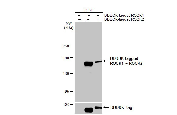
Non-transfected (–) and transfected (+) 293T whole cell extract (30 μg) were separated by 5% SDS-PAGE, and the membrane was blotted with ROCK1 + ROCK2 antibody [HL1632] (GTX637125) diluted at 1:5000. The HRP-conjugated anti-rabbit IgG antibody (GTX213110-01) was used to detect the primary antibody.
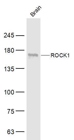
![Various whole cell extracts (30 μg) were separated by 5% SDS-PAGE, and the membrane was blotted with ROCK1 + ROCK2 antibody [HL1632] (GTX637125) diluted at 1:1000. The HRP-conjugated anti-rabbit IgG antibody (GTX213110-01) was used to detect the primary antibody.](https://www.genetex.com/upload/website/prouct_img/normal/GTX637125/GTX637125_45159_20230908_WB_23091319_348.webp "Various whole cell extracts (30 μg) were separated by 5% SDS-PAGE, and the membrane was blotted with ROCK1 + ROCK2 antibody [HL1632] (GTX637125) diluted at 1:1000. The HRP-conjugated anti-rabbit IgG antibody (GTX213110-01) was used to detect the primary antibody.")
![ROCK1 + ROCK2 antibody [HL1632] detects ROCK1 + ROCK2 protein at cytoplasm by immunohistochemical analysis. Sample: Paraffin-embedded human lung cancer. ROCK1 + ROCK2 stained by ROCK1 + ROCK2 antibody [HL1632] (GTX637125) diluted at 1:100. Antigen Retrieval: Citrate buffer, pH 6.0, 15 min](https://www.genetex.com/upload/website/prouct_img/normal/GTX637125/GTX637125_T-44739_20230925_IHC-P_23100319_384.webp "ROCK1 + ROCK2 antibody [HL1632] detects ROCK1 + ROCK2 protein at cytoplasm by immunohistochemical analysis. Sample: Paraffin-embedded human lung cancer. ROCK1 + ROCK2 stained by ROCK1 + ROCK2 antibody [HL1632] (GTX637125) diluted at 1:100. Antigen Retrieval: Citrate buffer, pH 6.0, 15 min")
![ROCK1 + ROCK2 antibody [HL1632] detects ROCK1 + ROCK2 protein at Golgi apparatus and cytoplasm by immunofluorescent analysis. Sample: HeLa cells were fixed in 4% paraformaldehyde at RT for 15 min. Green: ROCK1 + ROCK2 stained by ROCK1 + ROCK2 antibody [HL1632] (GTX637125) diluted at 1:500. Red: alpha Tubulin, a cytoskeleton marker, stained by alpha Tubulin antibody [GT114] (GTX628802) diluted at 1:1000. Blue: Fluoroshield with DAPI (GTX30920).](https://www.genetex.com/upload/website/prouct_img/normal/GTX637125/GTX637125_T-44739_20220826_ICC_IF_23102401_802.webp "ROCK1 + ROCK2 antibody [HL1632] detects ROCK1 + ROCK2 protein at Golgi apparatus and cytoplasm by immunofluorescent analysis. Sample: HeLa cells were fixed in 4% paraformaldehyde at RT for 15 min. Green: ROCK1 + ROCK2 stained by ROCK1 + ROCK2 antibody [HL1632] (GTX637125) diluted at 1:500. Red: alpha Tubulin, a cytoskeleton marker, stained by alpha Tubulin antibody [GT114] (GTX628802) diluted at 1:1000. Blue: Fluoroshield with DAPI (GTX30920).")
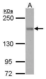
![Whole cell extract (30 μg) was separated by 5% SDS-PAGE, and the membrane was blotted with ROCK1 + ROCK2 antibody [HL1632] (GTX637125) diluted at 1:1000. The HRP-conjugated anti-rabbit IgG antibody (GTX213110-01) was used to detect the primary antibody.](https://www.genetex.com/upload/website/prouct_img/normal/GTX637125/GTX637125_45159_20231006_WB_R_23102401_237.webp "Whole cell extract (30 μg) was separated by 5% SDS-PAGE, and the membrane was blotted with ROCK1 + ROCK2 antibody [HL1632] (GTX637125) diluted at 1:1000. The HRP-conjugated anti-rabbit IgG antibody (GTX213110-01) was used to detect the primary antibody.")
![Whole cell extract (30 μg) was separated by 5% SDS-PAGE, and the membrane was blotted with ROCK1 + ROCK2 antibody [HL1632] (GTX637125) diluted at 1:1000. The HRP-conjugated anti-rabbit IgG antibody (GTX213110-01) was used to detect the primary antibody.](https://www.genetex.com/upload/website/prouct_img/normal/GTX637125/GTX637125_45159_20231006_WB_M_23102401_435.webp "Whole cell extract (30 μg) was separated by 5% SDS-PAGE, and the membrane was blotted with ROCK1 + ROCK2 antibody [HL1632] (GTX637125) diluted at 1:1000. The HRP-conjugated anti-rabbit IgG antibody (GTX213110-01) was used to detect the primary antibody.")
![ROCK1 + ROCK2 antibody [HL1632] detects ROCK1 + ROCK2 protein by immunohistochemical analysis. Sample: Paraffin-embedded rat tissues. ROCK1 + ROCK2 stained by ROCK1 + ROCK2 antibody [HL1632] (GTX637125) diluted at 1:200. Antigen Retrieval: Citrate buffer, pH 6.0, 15 min](https://www.genetex.com/upload/website/prouct_img/normal/GTX637125/GTX637125_45159_20231106_IHC-P_multiple_R_23111422_478.webp "ROCK1 + ROCK2 antibody [HL1632] detects ROCK1 + ROCK2 protein by immunohistochemical analysis. Sample: Paraffin-embedded rat tissues. ROCK1 + ROCK2 stained by ROCK1 + ROCK2 antibody [HL1632] (GTX637125) diluted at 1:200. Antigen Retrieval: Citrate buffer, pH 6.0, 15 min")
![ROCK1 + ROCK2 antibody [HL1632] detects ROCK1 + ROCK2 protein at cell membrane and cytoplasm by immunohistochemical analysis. Sample: Paraffin-embedded mouse colon. ROCK1 + ROCK2 stained by ROCK1 + ROCK2 antibody [HL1632] (GTX637125) diluted at 1:200. Antigen Retrieval: Citrate buffer, pH 6.0, 15 min](https://www.genetex.com/upload/website/prouct_img/normal/GTX637125/GTX637125_45159_20231106_IHC-P_M_23111422_109.webp "ROCK1 + ROCK2 antibody [HL1632] detects ROCK1 + ROCK2 protein at cell membrane and cytoplasm by immunohistochemical analysis. Sample: Paraffin-embedded mouse colon. ROCK1 + ROCK2 stained by ROCK1 + ROCK2 antibody [HL1632] (GTX637125) diluted at 1:200. Antigen Retrieval: Citrate buffer, pH 6.0, 15 min")
Non-transfected (–) and transfected (+) 293T whole cell extract (30 μg) were separated by 5% SDS-PAGE, and the membrane was blotted with ROCK1 + ROCK2 antibody [HL1632] (GTX637125) diluted at 1:5000. The HRP-conjugated anti-rabbit IgG antibody (GTX213110-01) was used to detect the primary antibody.
ROCK1 + ROCK2 antibody [HL1632]
GTX637125
ApplicationsImmunoFluorescence, Western Blot, ImmunoCytoChemistry, ImmunoHistoChemistry, ImmunoHistoChemistry Paraffin
Product group Antibodies
ReactivityHuman, Mouse, Rat
TargetROCK1
Overview
- SupplierGeneTex
- Product NameROCK1 + ROCK2 antibody [HL1632]
- Delivery Days Customer9
- Application Supplier NoteWB: 1:500-1:10000. *Optimal dilutions/concentrations should be determined by the researcher.Not tested in other applications.
- ApplicationsImmunoFluorescence, Western Blot, ImmunoCytoChemistry, ImmunoHistoChemistry, ImmunoHistoChemistry Paraffin
- CertificationResearch Use Only
- ClonalityMonoclonal
- Clone IDHL1632
- Concentration1 mg/ml
- ConjugateUnconjugated
- Gene ID6093
- Target nameROCK1
- Target descriptionRho associated coiled-coil containing protein kinase 1
- Target synonymsP160ROCK, ROCK-I, rho-associated protein kinase 1, p160 ROCK-1, renal carcinoma antigen NY-REN-35
- HostRabbit
- IsotypeIgG
- Protein IDO75116
- Protein NameRho-associated protein kinase 2
- ReactivityHuman, Mouse, Rat
- Storage Instruction-20°C or -80°C,2°C to 8°C
- UNSPSC12352203
Datasheet
Related products
Product group Antibodies
Anti-ROCK1 Antibody144-01008
ApplicationsImmunoPrecipitation, Western Blot, ImmunoHistoChemistry
ReactivityHuman, Mouse, Rat
TargetROCK1
- SizePrice

Product group Antibodies
Anti-ROCK1 Antibody Picoband(r)A00722-4-CARRIER-FREE
ApplicationsFlow Cytometry, Western Blot, ELISA
ReactivityHuman, Monkey, Mouse, Rat
TargetROCK1
- SizePrice
Product group Antibodies
ROCK1 antibodyGTX125921
ApplicationsWestern Blot
ReactivityHuman
TargetROCK1
- SizePrice
![Various whole cell extracts (30 μg) were separated by 5% SDS-PAGE, and the membrane was blotted with ROCK1 antibody [N1N2], N-term (GTX113266) diluted at 1:1000. The HRP-conjugated anti-rabbit IgG antibody (GTX213110-01) was used to detect the primary antibody.](https://www.genetex.com/upload/website/prouct_img/normal/GTX113266/GTX113266_44608_20220311_WB_w_23060501_218.webp)
Product group Antibodies
References
ROCK1 antibody [N1N2], N-termGTX113266
ApplicationsImmunoFluorescence, ImmunoPrecipitation, Western Blot, ImmunoCytoChemistry, ImmunoHistoChemistry, ImmunoHistoChemistry Paraffin
ReactivityHuman, Mouse, Rat
TargetROCK1
- SizePrice
![WB analysis of HEK293 (1) and ROCK1 (AA: 403-610)-hIgGFc transfected HEK293 (2) cell lysate using GTX60535 ROCK1 antibody [1H4].](https://www.genetex.com/upload/website/prouct_img/normal/GTX60535/GTX60535_20170912_WB_w_23061123_987.webp)
Product group Antibodies
ROCK1 antibody [1H4]GTX60535
ApplicationsWestern Blot, ELISA
ReactivityHuman
TargetROCK1
- SizePrice
![ROCK1 + ROCK2 antibody [GT261] detects ROCK1 protein by Western blot analysis. A. 30 μg 293T whole cell lysate/extract B. 30 μg HeLa whole cell lysate/extract C. 30 μg HepG2 whole cell lysate/extract 5 % SDS-PAGE ROCK1 + ROCK2 antibody [GT261] (GTX629971) dilution: 1:1000](https://www.genetex.com/upload/website/prouct_img/normal/GTX629971/GTX629971_41484_WB_w_23061202_231.webp)
Product group Antibodies
ROCK1 + ROCK2 antibody [GT261]GTX629971
ApplicationsWestern Blot
ReactivityHuman, Mouse, Rat
TargetROCK1
- SizePrice
![Various whole cell extracts (30 μg) were separated by 5% SDS-PAGE, and the membranes were blotted with ROCK1 + ROCK2 antibody [GT464] (GTX629972) diluted at 1:1000 and competitor's antibody diluted at 1:1000. The HRP-conjugated anti-mouse IgG antibody (GTX213111-01) was used to detect the primary antibody. *The competitor is not affiliated with GeneTex and does not endorse this product.](https://www.genetex.com/upload/website/prouct_img/normal/GTX629972/GTX629972_41484_20200117_WB_competitor_watermark_w_23061202_820.webp)
Product group Antibodies
ROCK1 + ROCK2 antibody [GT464]GTX629972
ApplicationsWestern Blot
ReactivityHuman, Mouse, Rat
TargetROCK1
- SizePrice
Product group Antibodies
ROCK1 Polyclonal AntibodyCAC15747
ApplicationsImmunoFluorescence, Western Blot, ELISA, ImmunoHistoChemistry
TargetROCK1
- SizePrice
Product group Antibodies
References
ROCK1 Polyclonal AntibodyBS-1166R
ApplicationsImmunoFluorescence, Western Blot, ELISA, ImmunoCytoChemistry, ImmunoHistoChemistry, ImmunoHistoChemistry Frozen, ImmunoHistoChemistry Paraffin
ReactivityHuman, Mouse, Rabbit, Rat
TargetROCK1
- SizePrice
Product group Antibodies
Anti-Rock-1 AntibodyA95593
ApplicationsWestern Blot, ELISA, ImmunoHistoChemistry
ReactivityHuman, Mouse, Rat
- SizePrice