

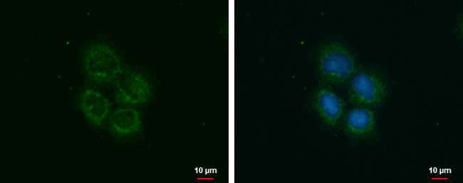
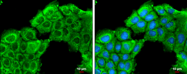
ROCK2 antibody detects ROCK2 protein at cytoplasm by immunofluorescent analysis. Sample: A431 cells were fixed in ice-cold MeOH for 5 min. Green: ROCK2 protein stained by ROCK2 antibody (GTX102619) diluted at 1:500. Blue: Hoechst 33342 staining.
 and transfected (+) 293T whole cell extracts (30 μg) were separated by 5% SDS-PAGE, and the membrane was blotted with ROCK2 antibody (GTX102619) diluted at 1:2000.")
 dilution: 1:1000")
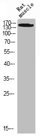
 was separated by 5% SDS-PAGE, and the membrane was blotted with ROCK2 antibody (GTX102619) diluted at 1:1000. The HRP-conjugated anti-rabbit IgG antibody (GTX213110-01) was used to detect the primary antibody, and the signal was developed with Trident ECL plus-Enhanced.")
 dilution: 1:1000")
 were separated by 5% SDS-PAGE, and the membranes were blotted with ROCK2 antibody (GTX102619) diluted at 1:1000 and competitor's antibody (sc-1851) diluted at 1:100. The HRP-conjugated anti-rabbit IgG antibody (GTX213110-01) was used to detect the primary antibody. *The competitor is not affiliated with GeneTex and does not endorse this product.")
![ROCK2 antibody detects ROCK2 protein expression by immunohistochemical analysis. Sample: Frozen-sectioned adult mouse cerebellum. Green: ROCK2 protein stained by ROCK2 antibody (GTX102619) diluted at 1:250. Red: NF-H, stained by NF-H antibody [GT114] (GTX634289) diluted at 1:500. Blue: Fluoroshield with DAPI (GTX30920).
Antigen Retrieval: Citrate buffer, pH 6.0, 10 min](https://www.genetex.com/upload/website/prouct_img/normal/GTX102619/GTX102619_41094_20170831_IHC-Fr_M_w_23060119_911.webp "ROCK2 antibody detects ROCK2 protein expression by immunohistochemical analysis. Sample: Frozen-sectioned adult mouse cerebellum. Green: ROCK2 protein stained by ROCK2 antibody (GTX102619) diluted at 1:250. Red: NF-H, stained by NF-H antibody [GT114] (GTX634289) diluted at 1:500. Blue: Fluoroshield with DAPI (GTX30920).
Antigen Retrieval: Citrate buffer, pH 6.0, 10 min")
 dilution: 1:500.
Antigen Retrieval: Trilogy? (EDTA based, pH 8.0) buffer, 15min")
 dilution: 1:1000")
. Western blot analysis was performed using ROCK2 antibody (GTX102619). EasyBlot anti-Rabbit IgG (GTX221666-01) was used as a secondary reagent.")
ROCK2 antibody detects ROCK2 protein at cytoplasm by immunofluorescent analysis. Sample: A431 cells were fixed in ice-cold MeOH for 5 min. Green: ROCK2 protein stained by ROCK2 antibody (GTX102619) diluted at 1:500. Blue: Hoechst 33342 staining.
ROCK2 antibody
GTX102619
ApplicationsImmunoFluorescence, ImmunoPrecipitation, Western Blot, ImmunoCytoChemistry, ImmunoHistoChemistry, ImmunoHistoChemistry Frozen, ImmunoHistoChemistry Paraffin
Product group Antibodies
ReactivityHuman, Mouse, Rat
TargetROCK2
Overview
- SupplierGeneTex
- Product NameROCK2 antibody
- Delivery Days Customer9
- Application Supplier NoteWB: 1:500-1:3000. ICC/IF: 1:100-1:1000. IHC-P: 1:100-1:1000. IHC-Fr: 1:100-1:1000. IP: 1:100-1:500. *Optimal dilutions/concentrations should be determined by the researcher.Not tested in other applications.
- ApplicationsImmunoFluorescence, ImmunoPrecipitation, Western Blot, ImmunoCytoChemistry, ImmunoHistoChemistry, ImmunoHistoChemistry Frozen, ImmunoHistoChemistry Paraffin
- CertificationResearch Use Only
- ClonalityPolyclonal
- Concentration0.4 mg/ml
- ConjugateUnconjugated
- Gene ID9475
- Target nameROCK2
- Target descriptionRho associated coiled-coil containing protein kinase 2
- Target synonymsROCK-II, rho-associated protein kinase 2, p164 ROCK-2, rho-associated, coiled-coil-containing protein kinase II
- HostRabbit
- IsotypeIgG
- Protein IDO75116
- Protein NameRho-associated protein kinase 2
- Scientific DescriptionThe protein encoded by this gene is a serine/threonine kinase that regulates cytokinesis, smooth muscle contraction, the formation of actin stress fibers and focal adhesions, and the activation of the c-fos serum response element. This protein, which is an isozyme of ROCK1 is a target for the small GTPase Rho. [provided by RefSeq]
- ReactivityHuman, Mouse, Rat
- Storage Instruction-20°C or -80°C,2°C to 8°C
- UNSPSC41116161
Datasheet
Related products
Product group Antibodies
ROCK2 AntibodyCSB-PA004018
ApplicationsWestern Blot, ELISA
ReactivityHuman, Monkey, Mouse, Rat
TargetROCK2
- SizePrice

Product group Antibodies
Anti-ROCK2 AntibodyA97284
ApplicationsWestern Blot, ELISA
ReactivityHuman, Mouse, Rat
- SizePrice

Product group Antibodies
Anti-ROCK2 Antibody Picoband(r)A01023-1-CARRIER-FREE
ApplicationsFlow Cytometry, ImmunoFluorescence, Western Blot, ELISA, ImmunoCytoChemistry
ReactivityHuman, Mouse, Rat
TargetROCK2
- SizePrice
Product group Antibodies

ROCK2 AntibodyLS-C831703
ApplicationsWestern Blot, ELISA
ReactivityHuman, Mouse, Rat
TargetROCK2
- SizePrice

Product group Antibodies
Anti-ROCK2 AntibodyHPA007459
ApplicationsWestern Blot, ImmunoHistoChemistry
ReactivityHuman, Mouse, Rat
TargetROCK2
- SizePrice
Product group Antibodies
ROCK2 Polyclonal AntibodyCAC14876
ApplicationsWestern Blot, ELISA
ReactivityMouse
TargetROCK2
- SizePrice
![Various whole cell extracts (30 μg) were separated by 5% SDS-PAGE, and the membranes were blotted with ROCK2 antibody [N3C1], Internal (GTX108247) diluted at 1:500 and competitor's antibody (sc-1851) diluted at 1:500. The HRP-conjugated anti-rabbit IgG antibody (GTX213110-01) was used to detect the primary antibody. *The competitor is not affiliated with GeneTex and does not endorse this product.](https://www.genetex.com/upload/website/prouct_img/normal/GTX108247/GTX108247_39735_20171222_WB_competitor_watermark_w_23060120_779.webp)
Product group Antibodies
ROCK2 antibody [N3C1], InternalGTX108247
ApplicationsImmunoFluorescence, Western Blot, ImmunoCytoChemistry, ImmunoHistoChemistry, ImmunoHistoChemistry Paraffin
ReactivityHuman, Mouse, Rat
TargetROCK2
- SizePrice
Product group Antibodies
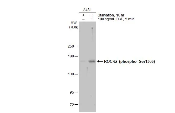
ROCK2 (phospho Ser1366) antibodyGTX122651
ApplicationsImmunoFluorescence, Western Blot, ImmunoCytoChemistry, ImmunoHistoChemistry, ImmunoHistoChemistry Frozen, ImmunoHistoChemistry Paraffin, Other Application
ReactivityHuman, Mouse, Rat
TargetROCK2
- SizePrice
Product group Antibodies
ROCK2 antibodyGTX122652
ApplicationsImmunoFluorescence, ImmunoPrecipitation, Western Blot, ImmunoCytoChemistry, ImmunoHistoChemistry, ImmunoHistoChemistry Paraffin
ReactivityHuman, Mouse
TargetROCK2
- SizePrice