

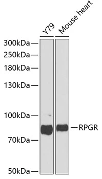
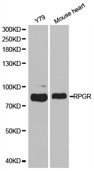
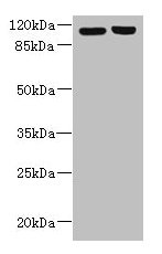
WB analysis of various sample lysates using GTX32845 RPGR antibody. Dilution : 1:1000 Loading : 25μg per lane
WB analysis of various sample lysates using GTX32845 RPGR antibody. Dilution : 1:1000 Loading : 25μg per lane
RPGR antibody
GTX32845
ApplicationsWestern Blot
Product group Antibodies
ReactivityHuman, Mouse
TargetRPGR
Overview
- SupplierGeneTex
- Product NameRPGR antibody
- Delivery Days Customer9
- Application Supplier NoteWB: 1:500 - 1:2000. *Optimal dilutions/concentrations should be determined by the researcher.Not tested in other applications.
- ApplicationsWestern Blot
- CertificationResearch Use Only
- ClonalityPolyclonal
- ConjugateUnconjugated
- Gene ID6103
- Target nameRPGR
- Target descriptionretinitis pigmentosa GTPase regulator
- Target synonymsCOD1, CORDX1, CRD, PCDX, RP15, RP3, XLRP3, orf15, X-linked retinitis pigmentosa GTPase regulator, retinitis pigmentosa 15, retinitis pigmentosa 3 GTPase regulator
- HostRabbit
- IsotypeIgG
- Protein IDQ92834
- Protein NameX-linked retinitis pigmentosa GTPase regulator
- Scientific DescriptionThis gene encodes a protein with a series of six RCC1-like domains (RLDs), characteristic of the highly conserved guanine nucleotide exchange factors. The encoded protein is found in the Golgi body and interacts with RPGRIP1. This protein localizes to the outer segment of rod photoreceptors and is essential for their viability. Mutations in this gene have been associated with X-linked retinitis pigmentosa (XLRP). Multiple alternatively spliced transcript variants that encode different isoforms of this gene have been reported, but the full-length natures of only some have been determined. [provided by RefSeq, Dec 2008]
- ReactivityHuman, Mouse
- Storage Instruction-20°C or -80°C,2°C to 8°C
- UNSPSC41116161
Datasheet
Related products
Product group Antibodies
Anti-RPGR AntibodyA30319
ApplicationsWestern Blot, ImmunoHistoChemistry
ReactivityHuman, Mouse, Rat
- SizePrice
Product group Antibodies
Anti-RPGR Antibody144-01844
ApplicationsWestern Blot
ReactivityHuman, Mouse
TargetRPGR
- SizePrice

Product group Antibodies
Anti-RPGR Antibody Picoband(r)A01522-CARRIER-FREE
ApplicationsWestern Blot, ELISA
ReactivityRat
TargetRPGR
- SizePrice
Product group Antibodies
RPGR AntibodyCSB-PA838814LA01HU
ApplicationsImmunoFluorescence, Western Blot, ELISA, ImmunoHistoChemistry
ReactivityHuman
TargetRPGR
- SizePrice
Product group Antibodies
Rpgr Polyclonal AntibodyCAC09543
ApplicationsImmunoFluorescence, Western Blot, ELISA, ImmunoHistoChemistry
TargetRPGR
- SizePrice
Product group Antibodies

RPGR AntibodyLS-C331722
ApplicationsWestern Blot, ImmunoHistoChemistry
ReactivityHuman, Mouse, Rat
TargetRPGR
- SizePrice
Product group Antibodies
Anti-RPGR AntibodyHPA001593
ApplicationsImmunoHistoChemistry
ReactivityHuman
TargetRPGR
- SizePrice