

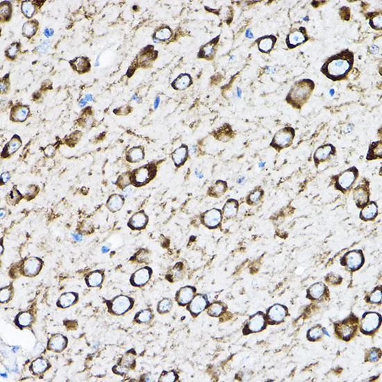
IHC-P analysis of mouse brain tissue using GTX66472 RPL17 antibody. Dilution : 1:100


IHC-P analysis of mouse brain tissue using GTX66472 RPL17 antibody. Dilution : 1:100
RPL17 antibody
GTX66472
ApplicationsImmunoFluorescence, Western Blot, ImmunoCytoChemistry, ImmunoHistoChemistry, ImmunoHistoChemistry Paraffin
Product group Antibodies
ReactivityHuman, Mouse, Rat
TargetRPL17
Overview
- SupplierGeneTex
- Product NameRPL17 antibody
- Delivery Days Customer9
- Application Supplier NoteWB: 1:200 - 1:2000. ICC/IF: 1:50 - 1:200. IHC-P: 1:50 - 1:200. *Optimal dilutions/concentrations should be determined by the researcher.Not tested in other applications.
- ApplicationsImmunoFluorescence, Western Blot, ImmunoCytoChemistry, ImmunoHistoChemistry, ImmunoHistoChemistry Paraffin
- CertificationResearch Use Only
- ClonalityPolyclonal
- ConjugateUnconjugated
- Gene ID6139
- Target nameRPL17
- Target descriptionribosomal protein L17
- Target synonymsDBA22, L17, PD-1, RPL23, uL22, large ribosomal subunit protein uL22, 60S ribosomal protein L17, 60S ribosomal protein L23, gene encoding putative NFkB activating protein
- HostRabbit
- IsotypeIgG
- Protein IDP18621
- Protein NameLarge ribosomal subunit protein uL22
- Scientific DescriptionRibosomes, the organelles that catalyze protein synthesis, consist of a small 40S subunit and a large 60S subunit. Together these subunits are composed of 4 RNA species and approximately 80 structurally distinct proteins. This gene encodes a ribosomal protein that is a component of the 60S subunit. The protein belongs to the L22P family of ribosomal proteins. It is located in the cytoplasm. This gene has been referred to as rpL23 because the encoded protein shares amino acid identity with ribosomal protein L23 from Halobacterium marismortui; however, its official symbol is RPL17. As is typical for genes encoding ribosomal proteins, there are multiple processed pseudogenes of this gene dispersed through the genome. Alternative splicing results in multiple transcript variants. Read-through transcription also exists between this gene and the neighboring downstream C18orf32 (chromosome 18 open reading frame 32) gene. [provided by RefSeq, Dec 2010]
- ReactivityHuman, Mouse, Rat
- Storage Instruction-20°C or -80°C,2°C to 8°C
- UNSPSC41116161
Datasheet
Related products
Product group Antibodies
RPL17 AntibodyCSB-PA003980
ApplicationsWestern Blot, ELISA, ImmunoHistoChemistry
ReactivityHuman, Monkey, Mouse, Rat
TargetRPL17
- SizePrice

Product group Antibodies
Anti-RPL17 AntibodyA95798
ApplicationsWestern Blot, ELISA, ImmunoHistoChemistry
ReactivityHuman, Mouse, Rat
- SizePrice

Product group Antibodies
Anti-RPL17 Antibody Picoband(r)A06980-2-CARRIER-FREE
ApplicationsFlow Cytometry, ImmunoFluorescence, Western Blot, ELISA, ImmunoCytoChemistry, ImmunoHistoChemistry
ReactivityHuman, Mouse, Rat
TargetRPL17
- SizePrice
Product group Antibodies
References

Goat anti-RPL17EB08028
ApplicationsWestern Blot, ELISA
ReactivityCanine, Human, Mouse, Rat
TargetRPL17
- SizePrice

Product group Antibodies
Anti-RPL17 AntibodyHPA043724
ApplicationsWestern Blot, ImmunoCytoChemistry, ImmunoHistoChemistry
ReactivityHuman
TargetRPL17
- SizePrice
Product group Antibodies

RPL17 / Ribosomal Protein L17 AntibodyLS-C334398
ApplicationsWestern Blot
ReactivityHuman, Mouse, Rat
TargetRPL17
- SizePrice
Product group Antibodies
RPL17 Polyclonal AntibodyCAC13983
ApplicationsWestern Blot, ELISA
TargetRPL17
- SizePrice
![Rat tissue extract (50 μg) was separated by 12% SDS-PAGE, and the membrane was blotted with RPL17 antibody [N1C3] (GTX101831) diluted at 1:500.](https://www.genetex.com/upload/website/prouct_img/normal/GTX101831/GTX101831_39918_20161020_WB_R_pancreas_w_23060100_919.webp)
Product group Antibodies
RPL17 antibody [N1C3]GTX101831
ApplicationsWestern Blot
ReactivityHuman, Rat
TargetRPL17
- SizePrice
![RPL17 antibody [N1C3-2] detects RPL17 protein at cytoplasm by immunofluorescent analysis. Sample: HeLa cells were fixed in 4% paraformaldehyde at RT for 15 min. Green: RPL17 protein stained by RPL17 antibody [N1C3-2] (GTX111934) diluted at 1:500. Red: alpha Tubulin, a cytoskeleton marker, stained by alpha Tubulin antibody [B-5-1-2] (GTX11304) diluted at 1:10000. Blue: Hoechst 33342 staining.](https://www.genetex.com/upload/website/prouct_img/normal/GTX111934/GTX111934_40086_20150410_IFA_w_23060500_218.webp)
Product group Antibodies
RPL17 antibody [N1C3-2]GTX111934
ApplicationsImmunoFluorescence, Western Blot, ImmunoCytoChemistry, ImmunoHistoChemistry, ImmunoHistoChemistry Paraffin
ReactivityHuman, Rat
TargetRPL17
- SizePrice