

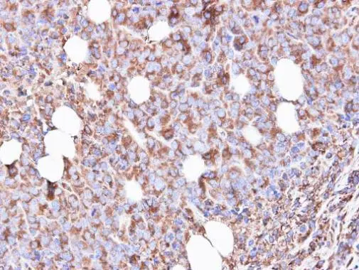
Immunohistochemical analysis of paraffin-embedded SAS xenograft, using RPS3(GTX103964) antibody at 1:500 dilution.
Antigen Retrieval: Trilogy? (EDTA based, pH 8.0) buffer, 15min
 of methanol-fixed HeLa, using RPS3(GTX103964) antibody (Green) at 1:500 dilution. Alpha-tubulin filaments were labeled with GTX11304 (Red) at 1:2000.")
 diluted at 1:500. Antigen Retrieval: Citrate buffer, pH 6.0, 15 min")
 diluted at 1:500.
Antigen Retrieval: Citrate buffer, pH 6.0, 15 min")
 diluted at 1:500.
Antigen Retrieval: Citrate buffer, pH 6.0, 15 min")
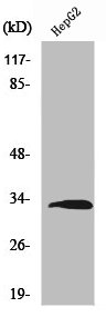
 were separated by 12% SDS-PAGE, and the membrane was blotted with RPS3 antibody (GTX103964) diluted at 1:500. The HRP-conjugated anti-rabbit IgG antibody (GTX213110-01) was used to detect the primary antibody.")
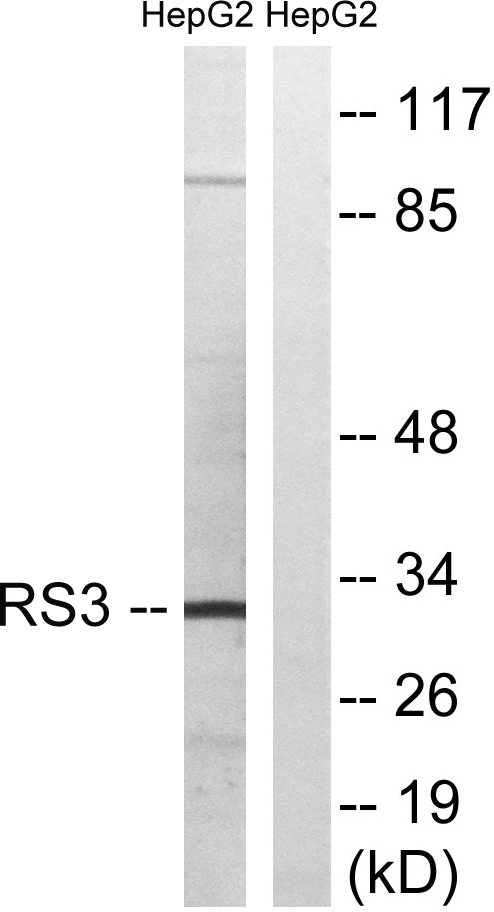
 were separated by 12% SDS-PAGE, and the membrane was blotted with RPS3 antibody (GTX103964) diluted at 1:500. The HRP-conjugated anti-rabbit IgG antibody (GTX213110-01) was used to detect the primary antibody. Corresponding RNA expression data are based on Human Protein Atlas program.")
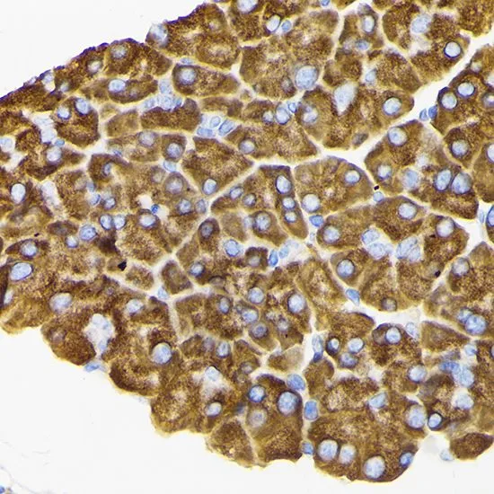
Immunohistochemical analysis of paraffin-embedded SAS xenograft, using RPS3(GTX103964) antibody at 1:500 dilution.
Antigen Retrieval: Trilogy? (EDTA based, pH 8.0) buffer, 15min
RPS3 antibody
GTX103964
ApplicationsImmunoFluorescence, ImmunoPrecipitation, Western Blot, ImmunoCytoChemistry, ImmunoHistoChemistry, ImmunoHistoChemistry Paraffin
Product group Antibodies
ReactivityHuman, Mouse, Rat
TargetRPS3
Overview
- SupplierGeneTex
- Product NameRPS3 antibody
- Delivery Days Customer9
- Application Supplier NoteWB: 1:500-1:3000. ICC/IF: 1:100-1:1000. IHC-P: 1:100-1:1000. *Optimal dilutions/concentrations should be determined by the researcher.Not tested in other applications.
- ApplicationsImmunoFluorescence, ImmunoPrecipitation, Western Blot, ImmunoCytoChemistry, ImmunoHistoChemistry, ImmunoHistoChemistry Paraffin
- CertificationResearch Use Only
- ClonalityPolyclonal
- Concentration1.38 mg/ml
- ConjugateUnconjugated
- Gene ID6188
- Target nameRPS3
- Target descriptionribosomal protein S3
- Target synonymsS3, uS3, small ribosomal subunit protein uS3, 40S ribosomal protein S3, IMR-90 ribosomal protein S3
- HostRabbit
- IsotypeIgG
- Protein IDP23396
- Protein NameSmall ribosomal subunit protein uS3
- Scientific DescriptionRibosomes, the organelles that catalyze protein synthesis, consist of a small 40S subunit and a large 60S subunit. Together these subunits are composed of 4 RNA species and approximately 80 structurally distinct proteins. This gene encodes a ribosomal protein that is a component of the 40S subunit, where it forms part of the domain where translation is initiated. The protein belongs to the S3P family of ribosomal proteins. Studies of the mouse and rat proteins have demonstrated that the protein has an extraribosomal role as an endonuclease involved in the repair of UV-induced DNA damage. The protein appears to be located in both the cytoplasm and nucleus but not in the nucleolus. Higher levels of expression of this gene in colon adenocarcinomas and adenomatous polyps compared to adjacent normal colonic mucosa have been observed. This gene is co-transcribed with the small nucleolar RNA genes U15A and U15B, which are located in its first and fifth introns, respectively. As is typical for genes encoding ribosomal proteins, there are multiple processed pseudogenes of this gene dispersed through the genome. [provided by RefSeq]
- ReactivityHuman, Mouse, Rat
- Storage Instruction-20°C or -80°C,2°C to 8°C
- UNSPSC41116161
Datasheet
Related products
Product group Antibodies
RPS3 AntibodyCSB-PA004002
ApplicationsWestern Blot, ELISA, ImmunoHistoChemistry
ReactivityHuman, Mouse, Rat
TargetRPS3
- SizePrice

Product group Antibodies
Anti-RPS3 Antibody Picoband(r)A01542-2-CARRIER-FREE
ApplicationsFlow Cytometry, ImmunoFluorescence, Western Blot, ImmunoCytoChemistry, ImmunoHistoChemistry
ReactivityHuman, Mouse, Rat
TargetRPS3
- SizePrice
Product group Antibodies
Anti-RPS3 AntibodyA96124
ApplicationsWestern Blot, ELISA, ImmunoHistoChemistry
ReactivityHuman, Mouse, Rat
- SizePrice
Product group Antibodies

RPS3 / Ribosomal Protein S3 AntibodyLS-C770370
ApplicationsWestern Blot, ELISA, ImmunoHistoChemistry
ReactivityHuman, Mouse, Rat
TargetRPS3
- SizePrice

Product group Antibodies
Anti-RPS3 AntibodyHPA063339
ApplicationsWestern Blot, ImmunoCytoChemistry, ImmunoHistoChemistry
ReactivityHuman
TargetRPS3
- SizePrice
Product group Antibodies
anti-Ribosomal Protein S3 (human), pAbAG-25A-0077
ApplicationsImmunoPrecipitation, Western Blot, ELISA, ImmunoCytoChemistry, ImmunoHistoChemistry
ReactivityHuman
TargetRPS3
- SizePrice
Product group Antibodies
RPS3 Recombinant AntibodyBSM-61209R
ApplicationsImmunoFluorescence, ImmunoCytoChemistry, ImmunoHistoChemistry, ImmunoHistoChemistry Frozen, ImmunoHistoChemistry Paraffin
TargetRPS3
- SizePrice
Product group Antibodies
RPS3 antibodyGTX54720
ApplicationsImmunoFluorescence, Western Blot, ImmunoCytoChemistry, ImmunoHistoChemistry, ImmunoHistoChemistry Paraffin
ReactivityHuman, Mouse, Rat
TargetRPS3
- SizePrice
Product group Antibodies
Anti-RPS3 Antibody144-02533
ApplicationsWestern Blot, ImmunoHistoChemistry
ReactivityHuman, Mouse, Rat
TargetRPS3
- SizePrice