

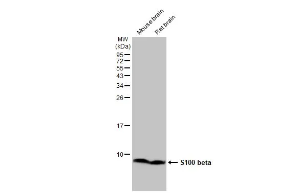
Various tissue extracts (50 μg) were separated by 15% SDS-PAGE, and the membrane was blotted with S100 beta antibody [HL2228] (GTX638273) diluted at 1:1000. The HRP-conjugated anti-rabbit IgG antibody (GTX213110-01) was used to detect the primary antibody.
![S100 beta antibody [HL2228] detects S100 beta protein by immunohistochemical analysis. Sample: Paraffin-embedded mouse eye. Green: S100 beta stained by S100 beta antibody [HL2228] (GTX638273) diluted at 1:100. Red: beta Tubulin 3/ Tuj1, a Cytoskeleton marker, stained by beta Tubulin 3/ Tuj1 antibody [GT11710] (GTX631836) diluted at 1:500. Blue: Fluoroshield with DAPI (GTX30920). Antigen Retrieval: Citrate buffer, pH 6.0, 15 min](https://www.genetex.com/upload/website/prouct_img/normal/GTX638273/GTX638273_T-44956_20230325_IHC-P_M_1_23032819_702.webp "S100 beta antibody [HL2228] detects S100 beta protein by immunohistochemical analysis. Sample: Paraffin-embedded mouse eye. Green: S100 beta stained by S100 beta antibody [HL2228] (GTX638273) diluted at 1:100. Red: beta Tubulin 3/ Tuj1, a Cytoskeleton marker, stained by beta Tubulin 3/ Tuj1 antibody [GT11710] (GTX631836) diluted at 1:500. Blue: Fluoroshield with DAPI (GTX30920). Antigen Retrieval: Citrate buffer, pH 6.0, 15 min")
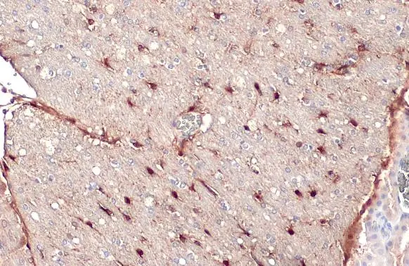
![S100 beta antibody [HL2228] detects S100 beta protein at cytoplasm and nucleus by immunohistochemical analysis. Sample: Paraffin-embedded mouse cerebellum. S100 beta stained by S100 beta antibody [HL2228] (GTX638273) diluted at 1:100. Antigen Retrieval: Citrate buffer, pH 6.0, 15 min](https://www.genetex.com/upload/website/prouct_img/normal/GTX638273/GTX638273_T-44956_20230325_IHC-P_M_23032819_347.webp "S100 beta antibody [HL2228] detects S100 beta protein at cytoplasm and nucleus by immunohistochemical analysis. Sample: Paraffin-embedded mouse cerebellum. S100 beta stained by S100 beta antibody [HL2228] (GTX638273) diluted at 1:100. Antigen Retrieval: Citrate buffer, pH 6.0, 15 min")
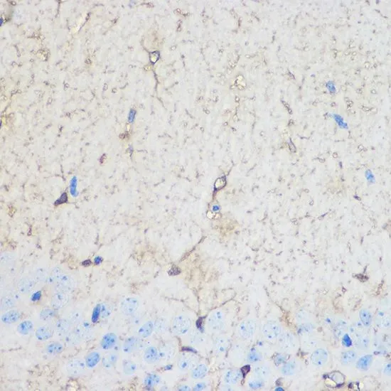
![S100 beta antibody [HL2228] detects S100 beta protein at cytoplasm and nucleus by immunohistochemical analysis. Sample: Paraffin-embedded mouse hippocampus. Green: S100 beta stained by S100 beta antibody [HL2228] (GTX638273) diluted at 1:100. Red: beta Tubulin 3/ Tuj1 , a Cytoskeleton marker, stained by beta Tubulin 3/ Tuj1 antibody [GT11710] (GTX631836) diluted at 1:500. Blue: Fluoroshield with DAPI (GTX30920). Antigen Retrieval: Citrate buffer, pH 6.0, 15 min](https://www.genetex.com/upload/website/prouct_img/normal/GTX638273/GTX638273_T-44956_20230414_IHC-P_M_23041719_463.webp "S100 beta antibody [HL2228] detects S100 beta protein at cytoplasm and nucleus by immunohistochemical analysis. Sample: Paraffin-embedded mouse hippocampus. Green: S100 beta stained by S100 beta antibody [HL2228] (GTX638273) diluted at 1:100. Red: beta Tubulin 3/ Tuj1 , a Cytoskeleton marker, stained by beta Tubulin 3/ Tuj1 antibody [GT11710] (GTX631836) diluted at 1:500. Blue: Fluoroshield with DAPI (GTX30920). Antigen Retrieval: Citrate buffer, pH 6.0, 15 min")
![S100 beta antibody [HL2228] detects S100 beta protein at cytoplasm and nucleus by immunohistochemical analysis. Sample: Paraffin-embedded rat hippocampus. Green: S100 beta stained by S100 beta antibody [HL2228] (GTX638273) diluted at 1:100. Red: beta Tubulin 3/ Tuj1 , a Cytoskeleton marker, stained by beta Tubulin 3/ Tuj1 antibody [GT11710] (GTX631836) diluted at 1:500. Blue: Fluoroshield with DAPI (GTX30920). Antigen Retrieval: Citrate buffer, pH 6.0, 15 min](https://www.genetex.com/upload/website/prouct_img/normal/GTX638273/GTX638273_T-44956_20230414_IHC-P_R_23041719_127.webp "S100 beta antibody [HL2228] detects S100 beta protein at cytoplasm and nucleus by immunohistochemical analysis. Sample: Paraffin-embedded rat hippocampus. Green: S100 beta stained by S100 beta antibody [HL2228] (GTX638273) diluted at 1:100. Red: beta Tubulin 3/ Tuj1 , a Cytoskeleton marker, stained by beta Tubulin 3/ Tuj1 antibody [GT11710] (GTX631836) diluted at 1:500. Blue: Fluoroshield with DAPI (GTX30920). Antigen Retrieval: Citrate buffer, pH 6.0, 15 min")
![Whole cell extract (30 μg) was separated by 15% SDS-PAGE, and the membrane was blotted with S100 beta antibody [HL2228] (GTX638273) diluted at 1:1000. The HRP-conjugated anti-rabbit IgG antibody (GTX213110-01) was used to detect the primary antibody.](https://www.genetex.com/upload/website/prouct_img/normal/GTX638273/GTX638273_45026_20230428_WB_23050223_378.webp "Whole cell extract (30 μg) was separated by 15% SDS-PAGE, and the membrane was blotted with S100 beta antibody [HL2228] (GTX638273) diluted at 1:1000. The HRP-conjugated anti-rabbit IgG antibody (GTX213110-01) was used to detect the primary antibody.")
![Non-transfected (–) and transfected (+) 293T whole cell extracts (30 μg) were separated by 15% SDS-PAGE, and the membrane was blotted with S100 beta antibody [HL2228] (GTX638273) diluted at 1:5000. The HRP-conjugated anti-rabbit IgG antibody (GTX213110-01) was used to detect the primary antibody.](https://www.genetex.com/upload/website/prouct_img/normal/GTX638273/GTX638273_45026_20230714_WB_B_23071822_329.webp "Non-transfected (–) and transfected (+) 293T whole cell extracts (30 μg) were separated by 15% SDS-PAGE, and the membrane was blotted with S100 beta antibody [HL2228] (GTX638273) diluted at 1:5000. The HRP-conjugated anti-rabbit IgG antibody (GTX213110-01) was used to detect the primary antibody.")
![S100 beta antibody [HL2228] detects S100 beta protein at glia by immunofluorescent analysis. Sample: DIV9 rat E18 primary cortical neuron and glia cells were fixed in 4% paraformaldehyde at RT for 15 min. Green: S100 beta stained by S100 beta antibody [HL2228] (GTX638273) diluted at 1:500. Blue: Fluoroshield with DAPI (GTX30920).](https://www.genetex.com/upload/website/prouct_img/normal/GTX638273/GTX638273_T-44956_20231222_ICC_IF_R_24011618_210.webp "S100 beta antibody [HL2228] detects S100 beta protein at glia by immunofluorescent analysis. Sample: DIV9 rat E18 primary cortical neuron and glia cells were fixed in 4% paraformaldehyde at RT for 15 min. Green: S100 beta stained by S100 beta antibody [HL2228] (GTX638273) diluted at 1:500. Blue: Fluoroshield with DAPI (GTX30920).")
![S100 beta antibody [HL2228] detects S100 beta protein by immunohistochemical analysis. Sample: Paraffin-embedded human glioblastoma. S100 beta stained by S100 beta antibody [HL2228] (GTX638273) diluted at 1:100. Antigen Retrieval: Citrate buffer, pH 6.0, 15 min](https://www.genetex.com/upload/website/prouct_img/normal/GTX638273/GTX638273_45026_20240626_IHC-P_24070822_755.webp "S100 beta antibody [HL2228] detects S100 beta protein by immunohistochemical analysis. Sample: Paraffin-embedded human glioblastoma. S100 beta stained by S100 beta antibody [HL2228] (GTX638273) diluted at 1:100. Antigen Retrieval: Citrate buffer, pH 6.0, 15 min")
![Indirect ELISA analysis was performed by coating the plate with recombinant E.coli expressed, full-length human S100 beta protein (nM) (46.73-0.73 nM). Coated protein was probed with S100 beta antibody [HL2228] (GTX638273) (1 μg/mL). Goat anti-rabbit IgG antibody (HRP) (GTX213110-01) (1:10000) was used to detect the bound primary antibody.](https://www.genetex.com/upload/website/prouct_img/normal/GTX638273/GTX638273_45026_20250418_ELISA_Indirect_25042420_363.webp "Indirect ELISA analysis was performed by coating the plate with recombinant E.coli expressed, full-length human S100 beta protein (nM) (46.73-0.73 nM). Coated protein was probed with S100 beta antibody [HL2228] (GTX638273) (1 μg/mL). Goat anti-rabbit IgG antibody (HRP) (GTX213110-01) (1:10000) was used to detect the bound primary antibody.")
Various tissue extracts (50 μg) were separated by 15% SDS-PAGE, and the membrane was blotted with S100 beta antibody [HL2228] (GTX638273) diluted at 1:1000. The HRP-conjugated anti-rabbit IgG antibody (GTX213110-01) was used to detect the primary antibody.
S100 beta antibody [HL2228]
GTX638273
ApplicationsImmunoFluorescence, Western Blot, ImmunoCytoChemistry, ImmunoHistoChemistry, ImmunoHistoChemistry Paraffin
Product group Antibodies
ReactivityHuman, Mouse, Rat
TargetS100B
Overview
- SupplierGeneTex
- Product NameS100 beta antibody [HL2228]
- Delivery Days Customer9
- Application Supplier NoteWB: 1:500-1:3000. *Optimal dilutions/concentrations should be determined by the researcher.Not tested in other applications.
- ApplicationsImmunoFluorescence, Western Blot, ImmunoCytoChemistry, ImmunoHistoChemistry, ImmunoHistoChemistry Paraffin
- CertificationResearch Use Only
- ClonalityMonoclonal
- Clone IDHL2228
- Concentration1 mg/ml
- ConjugateUnconjugated
- Gene ID6285
- Target nameS100B
- Target descriptionS100 calcium binding protein B
- Target synonymsNEF, S100, S100-B, S100beta, protein S100-B, S-100 calcium-binding protein, beta chain, S-100 protein subunit beta, S100 calcium-binding protein, beta (neural)
- HostRabbit
- IsotypeIgG
- Protein IDP04271
- Protein NameProtein S100-B
- Scientific DescriptionThe protein encoded by this gene is a member of the S100 family of proteins containing 2 EF-hand calcium-binding motifs. S100 proteins are localized in the cytoplasm and/or nucleus of a wide range of cells, and involved in the regulation of a number of cellular processes such as cell cycle progression and differentiation. S100 genes include at least 13 members which are located as a cluster on chromosome 1q21; however, this gene is located at 21q22.3. This protein may function in Neurite extension, proliferation of melanoma cells, stimulation of Ca2+ fluxes, inhibition of PKC-mediated phosphorylation, astrocytosis and axonal proliferation, and inhibition of microtubule assembly. Chromosomal rearrangements and altered expression of this gene have been implicated in several neurological, neoplastic, and other types of diseases, including Alzheimers disease, Downs syndrome, epilepsy, amyotrophic lateral sclerosis, melanoma, and type I diabetes. [provided by RefSeq, Jul 2008]
- ReactivityHuman, Mouse, Rat
- Storage Instruction-20°C or -80°C,2°C to 8°C
- UNSPSC12352203
Datasheet
Related products
Product group Antibodies

Anti-S100B [1C8]Ab02453-1.1
ApplicationsELISA
ReactivityHuman
TargetS100B
- SizePrice
Product group Antibodies
Anti-S100b Antibody130-00023
ApplicationsWestern Blot, ELISA
ReactivityHuman
- SizePrice

Product group Antibodies
References
ApplicationsImmunoFluorescence, ImmunoPrecipitation, Western Blot, ImmunoCytoChemistry, ImmunoHistoChemistry
ReactivityHuman, Mouse, Rat
TargetS100B
- SizePrice
Product group Antibodies
References
S100 beta antibodyGTX129573
ApplicationsImmunoFluorescence, Western Blot, ImmunoCytoChemistry, ImmunoHistoChemistry, ImmunoHistoChemistry Frozen, ImmunoHistoChemistry Paraffin
ReactivityFish, Human, Mammals, Mouse, Rat
TargetS100B
- SizePrice
![IHC-P analysis of human cerebrum (grey matter) tissue using GTX04404 S100 beta antibody [MSVA-490R] HistoMAX?. Strong ubiquitous S100 beta staining, except neirons.](https://www.genetex.com/upload/website/prouct_img/normal/GTX04404/GTX04404_20230728_IHC-P_107_23072722_172.webp)
Product group Antibodies
ApplicationsImmunoHistoChemistry, ImmunoHistoChemistry Paraffin
ReactivityHuman
TargetS100B
- SizePrice
![SDS-PAGE analysis of GTX04511 S100 beta antibody [4C4.9].](https://www.genetex.com/upload/website/prouct_img/normal/GTX04511/GTX04511_20230802_Image_23080120_268.webp)
Product group Antibodies
S100 beta antibody [4C4.9]GTX04511
ApplicationsFlow Cytometry, ImmunoFluorescence, Western Blot, ImmunoCytoChemistry, ImmunoHistoChemistry, ImmunoHistoChemistry Paraffin
ReactivityBovine, Human, Mouse, Rat
TargetS100B
- SizePrice
Product group Antibodies
References
S100 beta antibody [SH-B4]GTX11179
ApplicationsImmunoFluorescence, ELISA, ImmunoCytoChemistry, ImmunoHistoChemistry, ImmunoHistoChemistry Frozen, ImmunoHistoChemistry Paraffin
ReactivityBovine, Canine, Feline, Goat, Human, Mouse, Porcine, Rabbit, Rat, Sheep
TargetS100B
- SizePrice
Product group Antibodies
S100 beta antibodyGTX57757
ApplicationsImmunoFluorescence, ImmunoPrecipitation, Western Blot, ImmunoCytoChemistry, ImmunoHistoChemistry, ImmunoHistoChemistry Paraffin
ReactivityHuman, Mouse, Rat
TargetS100B
- SizePrice
![WB analysis of full-length S100B recombinant protein using GTX83327 S100 beta antibody [9A11B9].](https://www.genetex.com/upload/website/prouct_img/normal/GTX83327/GTX83327_20170912_WB_w_23061322_321.webp)
Product group Antibodies
S100 beta antibody [9A11B9]GTX83327
ApplicationsWestern Blot, ELISA, ImmunoHistoChemistry, ImmunoHistoChemistry Paraffin
ReactivityHuman
TargetS100B
- SizePrice