

Search results: Dengue virus
Product group Antibodies
References
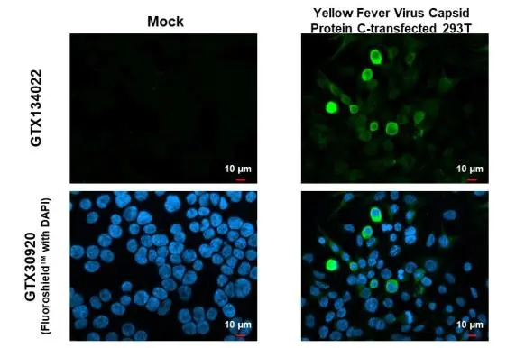
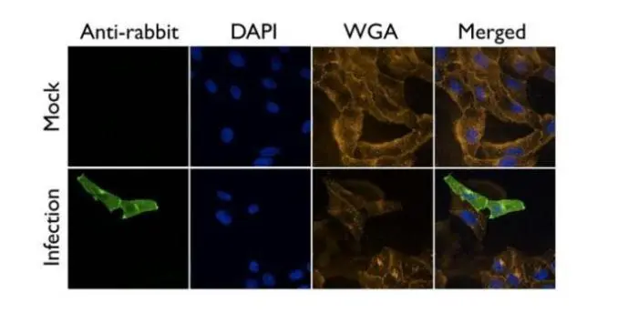
ApplicationsImmunoFluorescence, Western Blot, ImmunoCytoChemistry
ReactivityVirus
- SizePrice
Product group Antibodies
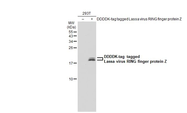
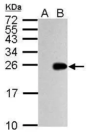
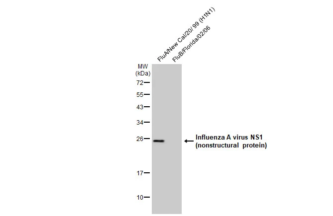
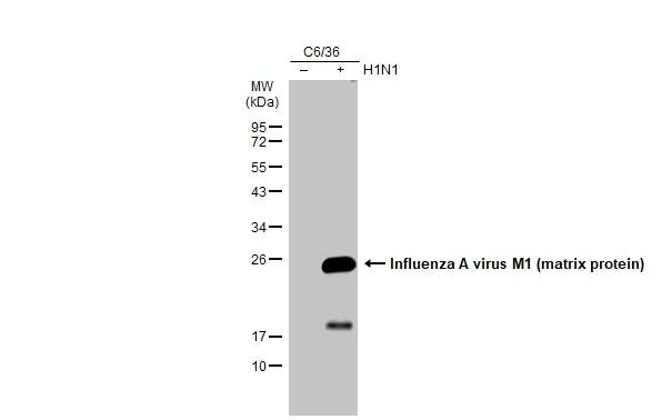
ApplicationsWestern Blot
ReactivityVirus
- SizePrice
Product group Antibodies
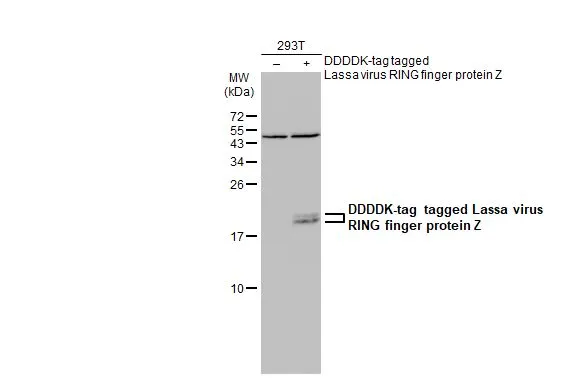
ApplicationsWestern Blot
ReactivityVirus
- SizePrice
Product group Antibodies
References
ApplicationsImmunoFluorescence, ImmunoPrecipitation, Western Blot, ELISA, ImmunoCytoChemistry, ImmunoHistoChemistry
ReactivityVirus
- SizePrice
Product group Antibodies
References
ApplicationsImmunoFluorescence, ImmunoPrecipitation, Western Blot, ELISA, ImmunoCytoChemistry, ImmunoHistoChemistry, Other Application
ReactivityVirus
- SizePrice
Product group Antibodies
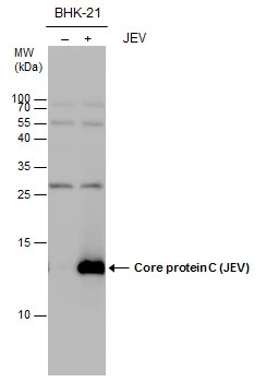
ApplicationsWestern Blot
ReactivityVirus
- SizePrice
Product group Antibodies
References
ApplicationsImmunoFluorescence, Western Blot, ImmunoCytoChemistry, ImmunoHistoChemistry, ImmunoHistoChemistry Frozen
ReactivityVirus
- SizePrice
Product group Antibodies
References
ApplicationsElectron Microscopy, ImmunoFluorescence, Western Blot, ImmunoCytoChemistry
ReactivityVirus
- SizePrice
Product group Antibodies
References
ApplicationsImmunoFluorescence, Western Blot, ImmunoCytoChemistry, ImmunoHistoChemistry, ImmunoHistoChemistry Paraffin
ReactivityVirus
- SizePrice

Product group Antibodies
ApplicationsWestern Blot
ReactivityVirus
- SizePrice
Product group Antibodies
References
ApplicationsWestern Blot, ELISA
ReactivityVirus
- SizePrice
Product group Antibodies
References
ApplicationsImmunoFluorescence, Western Blot, ELISA, ImmunoCytoChemistry, ImmunoHistoChemistry, ImmunoHistoChemistry Paraffin, Other Application
ReactivityVirus
- SizePrice
Didn't find what you were looking for?
Search through our product groups to find the right product
Back to overview